
![]()
Minor Burn Management
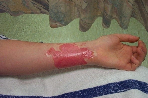
A 26 year-old female presented to the ED 20 minutes after spilling a hot sauce pan of soup onto her left forearm. The soup had just been brought to boil when the accident occurred. First aid was applied on arrival by placing the burn under under cold running water for 20 minutes.
Minor burns in the Emergency Department
- Minor burns are an important emergency department encounter because early treatment affecst the final outcome and keeping up-to-date with current treatment recommendations is challenging.
- They pose a high medico-legal risk to the clinician and there is the potential for psychological morbidity if scarring occurs.
- A study from Western Australia (Rea and Wood, 2005) looked at 10 months of minor burn injuries presenting to the regional burns unit. It showed that of the 227 patients, 45% sustained a scald, 19% a flame burn, 16% a contact burn, and 7.8% had a chemical burn. Of concern, 61% of the referred patients had not received appropriate first aid prior to referral.
Classification of Burns
Burns can be classified according to the body surface area that is involved:
- Minor Burns involve 10% TBSA or less
- Moderate Burns involve 11% to 20 of TBSA
- Major Burns involve 20% to 60% of TBSA
- Severe Burns involve >60% of TBSA
Types of Burns
There are 4 main types of burns that occur:
- Thermal
- Chemical
- Electrical
- Radiological
Classification of depth of burn injury
Burns are also classified according to the depth of injury:
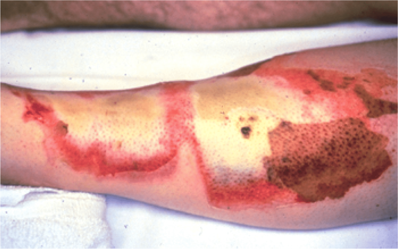
- Superficial — involves the epithelium: burns appear pink, red, painful, and generally take 7-10 days to heal. Sunburn is the classic example.
- Partial thickness — involves the epidermis and some dermis: appears mottled pink, painful, hairs intact, and generally take 2-3 weeks to heal. Scarring may occur, particularly if healing is delayed and skin grafts may be required.
- Full thickness — extends through the skin to deeper structures: appears black or white (eschar), leathery, produces no response to pain, and hairs are absent. Skin grafts are required.
Pathophysiology of burn injuries
The local response to a burn injury is described as consisting of three zones:
- Zone of coagulative necrosis results from the direct thermal injury at time of exposure.
- Zone of stasis borders the site of coagulation necrosis, there is a prominent inflammatory response and vascular reactivity that reduces blood flow. This reduction in flow occurs for the first 24-48 hours after the burn occurs.
- Zone of hyperaemia is the outermost area of the burn injury and is characterised by intense yet reversible vasodilatation and increased blood flow.
Thermal injuries trigger an intense local and systemic inflammatory response, with increased capillary permeability causing fluids and proteins to leak from the vascular space. This leakage can lead to oedema and hypovolaemia in extensive burns.
Emergency Department Management of Minor Thermal Burns
The 4 main aims of burns treatment are:
- Protection from the environment (infection)
- Temperature control (hypothermia)
- Fluid control (dehydration)
- Energy control (need for increased caloric intake)
First Aid
- Initial first aid should consist of cool running water (2-15°C), applied for a 20 minute duration. This should be performed as soon as possible but may be effective up to 3 hours after the burn occurred.
- The application of cool running water has been shown to significantly reduce soft tissue damage, hastens wound re-epithelialisation and reduces scaring.
- Ice should be avoided although effective at relieving pain, can cause a thermal burn in itself to the already damaged epithelial cells.
- All alternative therapies should be avoided as effectiveness has not been demonstrated in the literature. Do not smear the burn with butter!
- In Australia most prehospital provider service have adopted the use of Burnaid® dressings — a hydrocolloid type dressing that contains tea tree oil.
- They are marketed to relieve pain, infection and inflammation. However current evidence has shown no antibacterial effects against common burn pathogens and allergic contact dermatitis is a common adverse effect.
- It is amazing that these product are being used so frequently with very little evidence to support their use. They did not reduce scarring or inflammation in a porcine model although they are effective at cooling burns. They should be avoided in patients with over 20% TBSA due to the risk of hypothermia.
Cleaning burns
- Burns are at high risk of infection as breakdown of the epidermis denudes the body of a strong protective barrier against micro-organisms.
- All burns that are contaminated, or have been de-roofed or debrided, should be thoroughly irrigated.
- Choosing between tap water or saline as the irrigating fluid does not alter outcome or infection risk, although the goal of trying to maintain a sterile environment means most providers prefer to use saline.
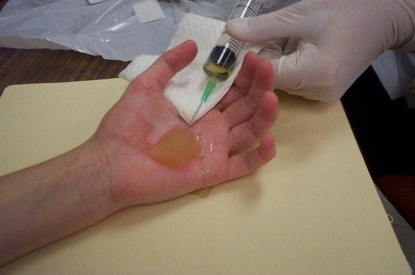
Blister Management
- This is a controversy that cannot be killed — should you leave blisters alone, aspirate them or de-roof them???
- There is limited evidence available to guide clinical practice, which is largely based on expert opinion.
- Leave the blisters alone?
- pros — they provides an barrier for the burn site against infection.
- cons — the underly damage to the epithelium cannot be visualised and the serous fluid in the blister may be impairing healing to the burn. The blister may impair joint functionality.
- Many specialists now recommend de-roofing and debriding the blister, so the underlying wound bed can be assessed and managed accordingly, and so that any non-adherent de-vitalised tissue is debrided.
- Follow local institutional guidelines or obtain specialist advice from your burns unit.
Dressings
- Choosing the correct dressing to manage these burns can be bewildering. Again, consulting local guidelines or obtaining specialist advice is wise.
- Although there are many dressings available on the market to treat burns, there is little evidence to support the use of one over the other.
- With appropriate wound management partial thickness burns should take around 10-12 days to heal.
- The majority of superficial burns do not require a dressing, and should heal nicely with good first aid and the application of emollient creams (e.g. sorbelene).
- Partial thickness and full thickness burns require dressings to aid healing. Types of dressing available are hydrocolloid, silicon nylon, antimicrobial, polyurethane film and biosynthetic dressings.
- The most common dressing I use on burns is the antimicrobial dressing Acticoat©, it contains the antimicrobial nano-crystalline silver.
- Silver has been shown have excellent antimicrobial effects and starts killing bacteria within 30mins of application and last up to 3 days as long as the dressing is kept wet.
- The dressing has a silver and a blue side — the blue side is placed onto the wound after the silver has to be activated with water for injection (do not use saline as it deactivates the silver). A second dressing needs to place over the to secure it, and allow for the dressing to retain moisture, occlusive dressings like Duoderm© works well for this. The dressing should be check daily to make sure it remains moist.
- Silver Sulphadiazine (SSD) creams have been commonly used in the past for the treatment of burns, however their effectiveness has been questioned.
- There is little evidence to suggest that SSD creams reduce bacterial infections in burns and local hypersensitivity reactions are common. Another downfall of SSD creams is they need to be removed and reapplied daily. This is inconvenient and expensive.
- Most burns units have stopped recommending the use of SSD creams
Analgesia
- Superficial and partial thickness burns are generally painful as nociceptors are intact.
- Simple analgesia like paracetamol and NSAIDs are generally effective for minor burns.
- In the acute period IV opiates may be required.
Antibiotics
- Antibiotics are not recommend as prophylaxis for minor burns.
- Appropriate first aid and ongoing sterile dressing changes decrease the risk of wound infection.
- If signs of infection are present, wound swabs should be taken to guide appropriate antibiotic use.
Tetanus
- Burns are high risk wounds for developing Clostridium tetani
- Tetanus status should be determined early and an ADT booster provided as required.
Follow up
- Follow up will depend on the patient’s location, access to a specialised burns clinic and the frequency of dressing changes required.
- Remember the burn will look worse 2-3 days after it occurs, so early follow up 48-72 hours after the injury should be arranged.
- Burns taking longer than 12-14days should be referred to the burns unit for assessment and to prevent scar formation.
Medico-legal Pitfalls
- Failure to suspect that non-accidental injury could be the cause of burns in children
- Not referring minor burns that are taking long time to heal, placing the patient at risk of scarring and psychosocial impairment.
- Burns will always look worse the next day, early follow up will detect changes in the severity of the burn.

References
- First Aid for Emergency Situations. ACLS Training Centre
- Bezuhly, M. Gomez, M. & Fish, J. (2004). Emergency department management of minor burn injuries in Ontario, Canada. Burns. 30, 160-164. PMID:15019126
- Burns Blister Management: Evidence Summary. (2009). Australasian Cochrane Centre
- Cuttle, L. & Kimble, RM. (2010). First aid treatment of burn injuries. Wound Practise and Research. 18(1),6-13.
- Ehrlich, A. et.al. (2005). Elderly patients discharged home from the emergency department with minor burns. Burns. 31, 717-720. PMID: 16039785
- Gomez, R. & Cancio, L. (2007). Management of Burn Wounds in the Emergency Department. Emergency Medicine Clinics of North America. 25, 135-146. PMID: 17400077
- Gravante, G. et.al. (2009). Nanoncrystalline silver: a systematic review of randomised trials conducted on burned patients and an evidence-based assessment of potential advantages over older silver formulations. Annals of Plastic Surgery. 63(2), 201-205. PMID: 19571738
- Hettiaratchy, S. Papini, R. & Dziewulski, P. (2005). ABC of Burns. Massachusetts: Blackwell Publishing
- Hussain, S.& Ferguson, C. (2009). Assessing the size of burns in patients- which method works best? www.bestbets.org
- Hussain, S. & Ferguson, C. (2005). Cooling as analgesia for burns. www.bestbets.org
- Hussain, S. & Ferguson, C. (2006). Silver Sulphadiazine cream in burns.www.bestbets.org
- Martin, R. (2007). Do Burnshield® dressings reduce the analgesia requirements and length of stay of for emergency patients. Australasian Emergency Nursing Journal. 208-209.
- Rea, S. Kuthubutheen, J. Fowler, B. & Wood, F. (2005). Burn first aid in Western Australia- Do healthcare workers have the knowledge? Burns. 1029-1034. PMID: 16308098
- Rea, S. & Wood, F. (2005). Minor burn injuries in adults presenting to regional burns unit in Western Australia: A prospective descriptive study. Burns. 1035-1040. PMID: 16289333
- Sargent, R. (2006). Management of Blisters in the Partial-Thickness Burn: An Integrative Research Review. Journal of Burn Care & Research. 27(1), 66-81.PMID: 16566539
- Shaw, J. & Dibble, C. (2006). Management of Burns Blisters. www.bestbets.org
- Singer, A. Brebbia, J. & Soroff, H. (2007). Management of local burn wounds in the ED. American Journal of Emergency Medicine. 25, 666-671. PMID: 17606093
- Singer, A. (2000). Thermal Burns: Rapid Assessment and Treatment. Emergency Medicine Practise. 2(9),1-20.
- Wasiak, J. Cleland, H. & Campbell, F. (2009). Dressings for superficial and partial thickness burns (Review).The Cochrane Collaboration
- Wasiak, J. et.al. (2009). The epidemiology of burn injuries in an Australian setting, 2000-2006. Burns. 35, 1124-1132. PMID: 19482430
Emergency nurse with ultra-keen interest in the realms of toxicology, sepsis, eLearning and the management of critical care in the Emergency Department | LinkedIn |