
![]()
Hyperkalaemia
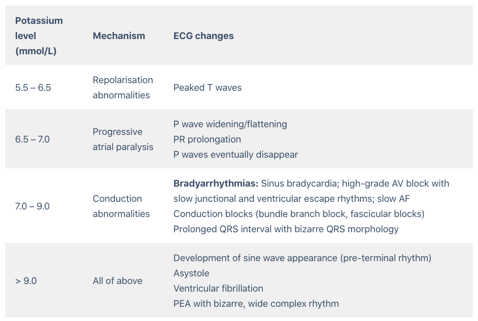
Hyperkalaemia is defined as a serum potassium level of > 5.2 mmol/L. ECG changes generally do not manifest until there is a moderate degree of hyperkalaemia (≥ 6.0 mmol/L). The earliest manifestation of hyperkalaemia is an increase in T wave amplitude.
ECG features of hyperkalaemia
- Peaked T waves
- P wave widening/flattening, PR prolongation
- Bradyarrhythmias: sinus bradycardia, high-grade AV block with slow junctional and ventricular escape rhythms, slow AF
- Conduction blocks (bundle branch block, fascicular blocks)
- QRS widening with bizarre QRS morphology
With worsening hyperkalaemia… (> 9.0 mmol/L):
- Development of sine wave appearance (pre-terminal rhythm)
- Ventricular fibrillation
- PEA with bizarre, wide complex rhythm
- Asystole
Note: Serum potassium level may not correlate closely with ECG changes. Patients with a relatively normal ECG can suffer sudden hyperkalaemia cardiac arrest. In any patient who has suffered a bradycardia PEA arrest, suspect and treat for hyperkalaemia.
An easy way to remember the usual order of ECG changes seen is by following the ECG trace logically – effects begin on the T wave and move forwards to the P wave / PR interval, and subsequently to the QRS complex with QRS widening and conduction blocks.
The push-pull effect
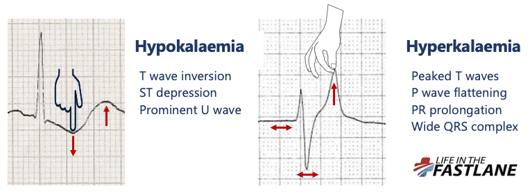
- Hypokalaemia creates the illusion that the T wave is “pushed down”, with resultant T-wave flattening/inversion, ST depression, and prominent U waves
- In hyperkalaemia, the T wave is “pulled upwards”, creating tall “tented” T waves, and stretching the remainder of the ECG to cause P wave flattening, PR prolongation, and QRS widening
Pathophysiology
Potassium is vital for regulating the normal electrical activity of the heart. Increased extracellular potassium reduces myocardial excitability, with depression of both pacemaking and conducting tissues.
Progressively worsening hyperkalaemia leads to suppression of impulse generation by the SA node and reduced conduction by the AV node and His-Purkinje system, resulting in bradycardia and conduction blocks and ultimately cardiac arrest.
| Degree of hyperkalaemia | Potassium level (mmol/L) |
| Mild | 5.3 – 6.0 |
| Moderate | 6.0 – 6.9 |
| Severe | ≥ 7.0 |
Handy Tips
Suspect hyperkalaemia in any patient with a new bradyarrhythmia or AV block, especially patients with renal failure, on haemodialysis, or taking any combination of ACE inhibitors, potassium-sparing diuretics and potassium supplements.
For an excellent review of the management of hyperkalaemia, check out this podcast by Scott Weingart.
ECG Examples
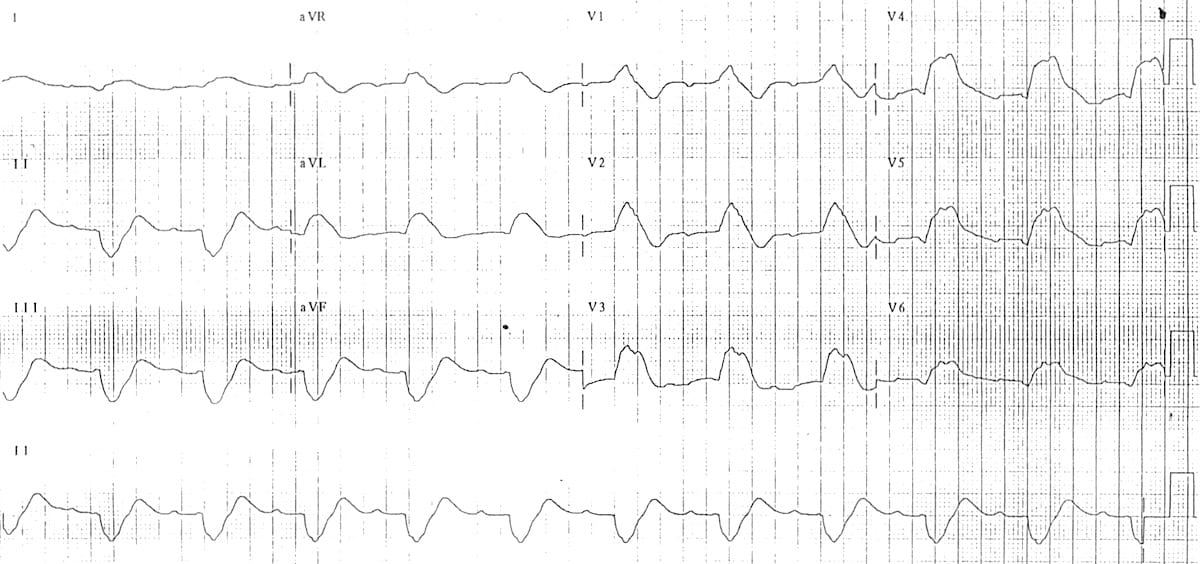
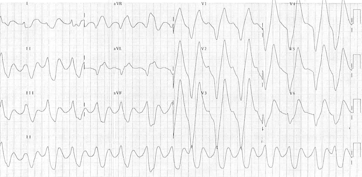
Example 1
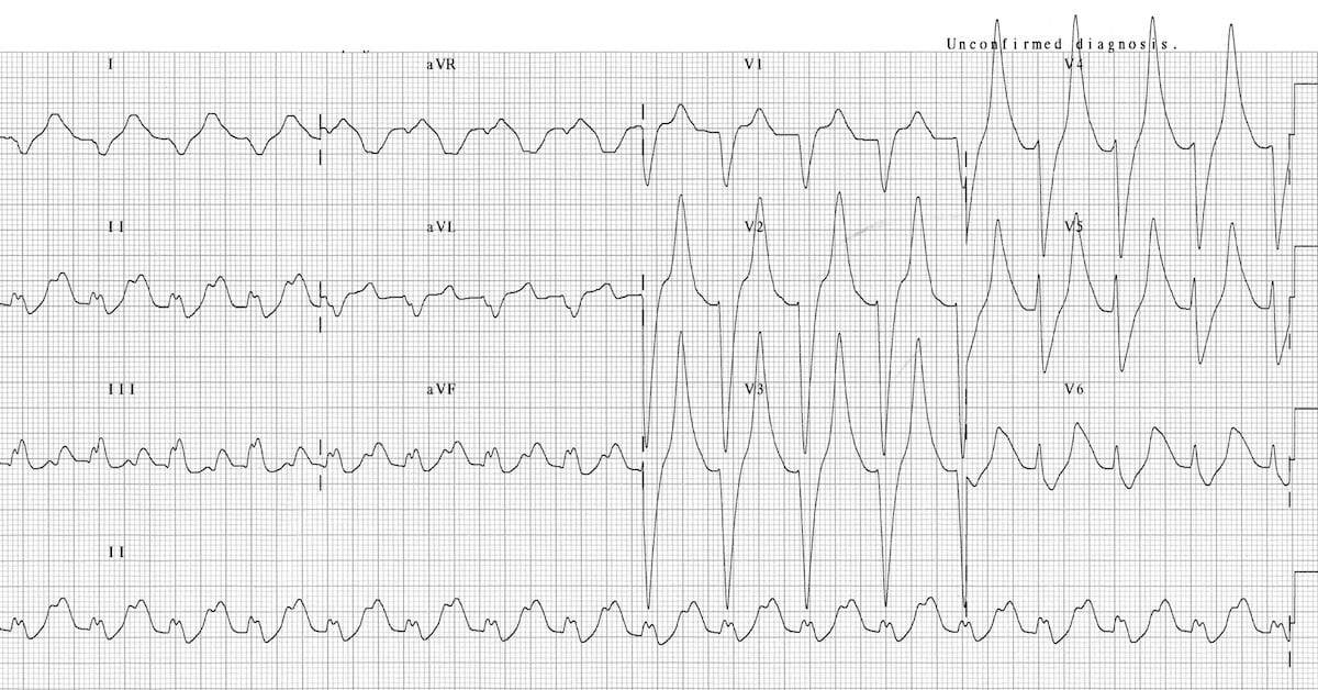
This ECG displays many of the features of hyperkalaemia:
- Prolonged PR interval.
- Broad, bizarre QRS complexes — these merge with both the preceding P wave and subsequent T wave.
- Peaked T waves.
This patient had a serum K+ of 9.3
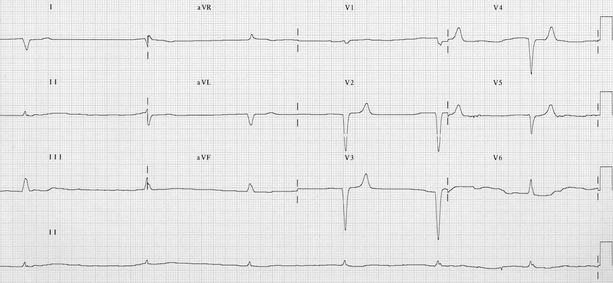
Example 2
Hyperkalaemia
- Tall, symmetrically peaked T waves.
This patient had a serum K+ of 7.0.
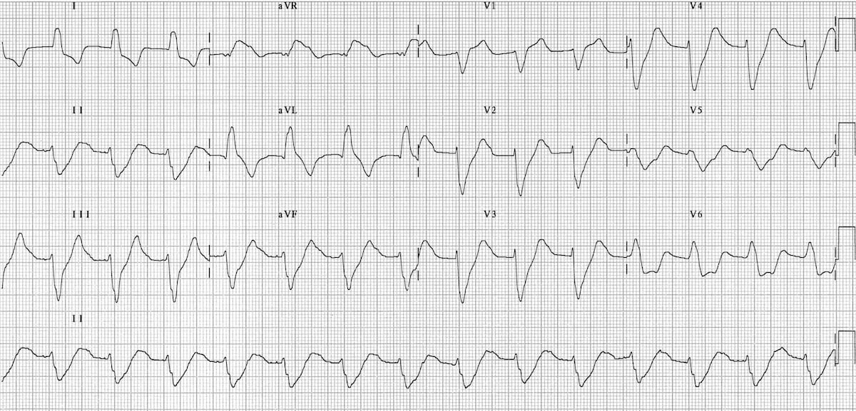
Example 3
Hyperkalaemia
- Long PR segment.
- Wide, bizarre QRS.
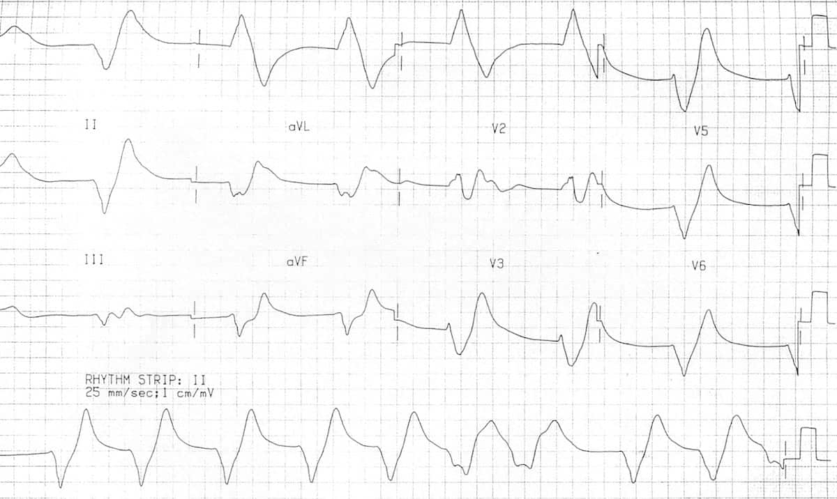
Example 4
Hyperkalaemia:
- Slow junctional rhythm.
- Intraventricular conduction delay.
- Peaked T waves.
Example 5
Hyperkalaemia:
- Broad complex rhythm with atypical LBBB morphology.
- Left axis deviation.
- Absent P waves.
Example 6
Hyperkalaemia:
- Sine wave appearance with severe hyperkalaemia (K+ 9.9 mEq/L).
Example 7
Hyperkalaemia:
- Huge peaked T waves.
- Sine wave appearance.
This patient had severe hyperkalaemia (K+ 9.0 mEq/L) secondary to rhabdomyolysis.
Related Topics
Advanced Reading
Online
- Wiesbauer F, Kühn P. ECG Mastery: Yellow Belt online course. Understand ECG basics. Medmastery
- Wiesbauer F, Kühn P. ECG Mastery: Blue Belt online course: Become an ECG expert. Medmastery
- Kühn P, Houghton A. ECG Mastery: Black Belt Workshop. Advanced ECG interpretation. Medmastery
- Rawshani A. Clinical ECG Interpretation ECG Waves
- Smith SW. Dr Smith’s ECG blog.
Textbooks
- Zimmerman FH. ECG Core Curriculum. 2023
- Mattu A, Berberian J, Brady WJ. Emergency ECGs: Case-Based Review and Interpretations, 2022
- Straus DG, Schocken DD. Marriott’s Practical Electrocardiography 13e, 2021
- Brady WJ, Lipinski MJ et al. Electrocardiogram in Clinical Medicine. 1e, 2020
- Mattu A, Tabas JA, Brady WJ. Electrocardiography in Emergency, Acute, and Critical Care. 2e, 2019
- Hampton J, Adlam D. The ECG Made Practical 7e, 2019
- Kühn P, Lang C, Wiesbauer F. ECG Mastery: The Simplest Way to Learn the ECG. 2015
- Grauer K. ECG Pocket Brain (Expanded) 6e, 2014
- Surawicz B, Knilans T. Chou’s Electrocardiography in Clinical Practice: Adult and Pediatric 6e, 2008
- Chan TC. ECG in Emergency Medicine and Acute Care 1e, 2004
LITFL Further Reading
- ECG Library Basics – Waves, Intervals, Segments and Clinical Interpretation
- ECG A to Z by diagnosis – ECG interpretation in clinical context
- ECG Exigency and Cardiovascular Curveball – ECG Clinical Cases
- 100 ECG Quiz – Self-assessment tool for examination practice
- ECG Reference SITES and BOOKS – the best of the rest
[cite]
ECG LIBRARY

MBBS (UWA) CCPU (RCE, Biliary, DVT, E-FAST, AAA) Adult/Paediatric Emergency Medicine Advanced Trainee in Melbourne, Australia. Special interests in diagnostic and procedural ultrasound, medical education, and ECG interpretation. Editor-in-chief of the LITFL ECG Library. Twitter: @rob_buttner
Emergency Physician in Prehospital and Retrieval Medicine in Sydney, Australia. He has a passion for ECG interpretation and medical education | ECG Library |
thank you very much for easily memorable explanation ; push-pull effect
[…] Hyperkalaemia […]