
![]()
Ultrasound Case 089
Presentation
A woman of 40 presents with intermittent RUQ pain and nausea. You try to find her gallbladder.
Describe and interpret these scans
IMAGE INTERPRETATION
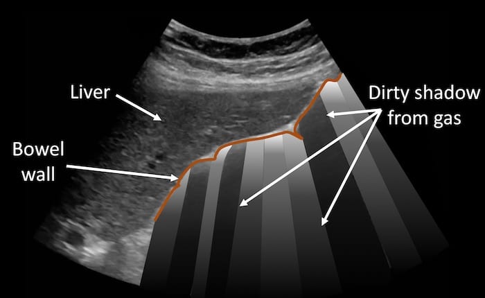
Image 1: Fanning through the long axis of the gallbladder fossa.
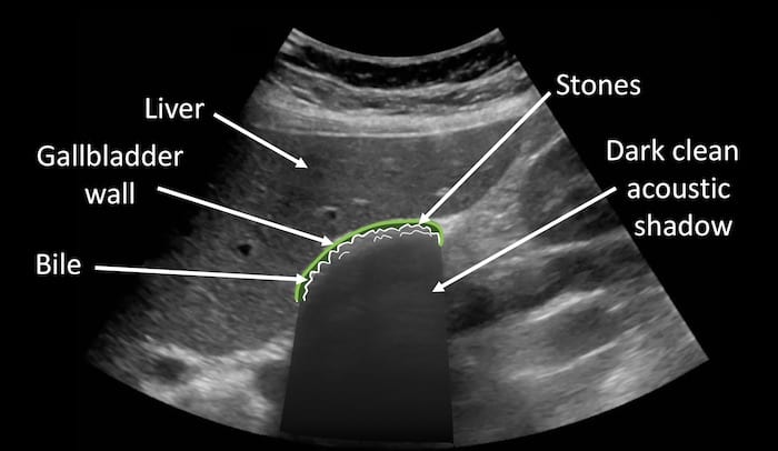
This lovely clip shows how difficult it can be to differentiate gas within the lumen of bowel from a gallbladder packed with tiny stones. The clip shows the liver edge covering gas filled bowel and then fans to show the wall-echo-shadow pattern typical of a gallbladder tightly packed with small stones.
CLINICAL CORRELATION
Cholelithiasis with the WES sign
Air and stones can appear similar on ultrasound.
- Air casts a dirty grey shadow.
- Stone casts a darker cleaner acoustic shadow.
Wall-echo-shadow (WES) sign: first there is the echogenic gallbladder wall, then a very thin rim of hypoechoic bile, then the echogenic surface may by innumerable small gallstones, and finally the dark and clean acoustic shadow.
[cite]
TOP 100 ULTRASOUND CASES

An Emergency physician based in Perth, Western Australia. Professionally my passion lies in integrating advanced diagnostic and procedural ultrasound into clinical assessment and management of the undifferentiated patient. Sharing hard fought knowledge with innovative educational techniques to ensure knowledge translation and dissemination is my goal. Family, wild coastlines, native forests, and tinkering in the shed fills the rest of my contented time. | SonoCPD | Ultrasound library | Top 100 | @thesonocave |