
![]()
Ultrasound Case 092
Presentation
A 38 year old patient with diabetes and end-stage renal failure presents with arm pain and sepsis. You are asked whether there is a drainable collection.
View 2: Longitudinal volar forearm
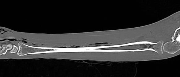
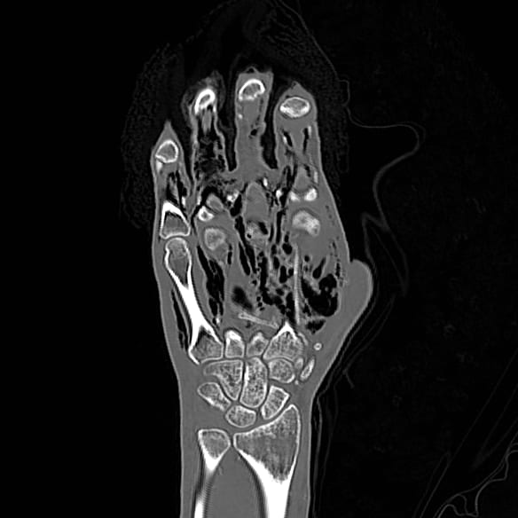
View 3 and 4: CT scans of the forearm and hand
Describe and interpret these scans
IMAGE INTERPRETATION
Image 1: Transverse mid left forearm with compression.
There is no obvious collection seen. You are searching for an abscess, usually hypoechoic with swirling internal debris on probe pressure. Your eye however is attracted to two echogenic foci. These have dirty posterior acoustic shadowing and you wonder if they could be air.
Image 2: Longitudinal mid forearm over the radius.
Here multiple foci of air coalesce in the mid facial plane (about 12.5 mm deep) obscuring deeper tissues with dirty shadowing.
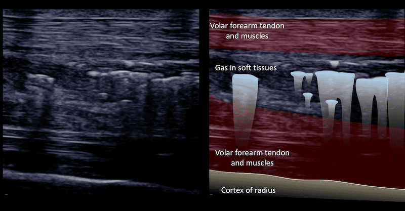
Image 4 & 5: CT scans of the forearm and hand showing extensive subcutaneous gas. This patient had necrotising fasciitis.
CLINICAL CORRELATION
Necrotising fasciitis
Gas in the soft tissues is not an infrequent finding. The typical picture is of small echobright locules with dirty posterior acoustic shadowing. Gas may be injected into the tissues, may spread from disruption of a gas filled organ, or may be formed by a gas forming organism.
[cite]
TOP 100 ULTRASOUND CASES