![]()

Abdominal Aortic Aneurysm (AAA)
Overview of Abdominal Aortic Aneurysm: presentation, risk factors, rupture risk, clinical features, investigations, emergency management, and surgical options

![]()
Overview of Abdominal Aortic Aneurysm: presentation, risk factors, rupture risk, clinical features, investigations, emergency management, and surgical options
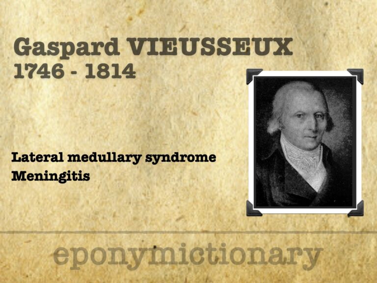
Swiss physician Gaspard Vieusseux (1746–1814) described cerebrospinal meningitis in 1805 and gave the first clinical account of lateral medullary syndrome.
Lemierre syndrome is infective thrombophlebitis of the internal jugular vein caused primarily by anaerobic organisms from a focus of oropharyngeal infection

Emergency Procedure: Today we tackle postpartum haemorrhage (PPH). Take a few deep breaths and your own pulse, then dive into the video

Adriaan van den Spiegel (1578–1625), Flemish anatomist; described Spigelian line, fascia, hernia, and liver lobe in his posthumous atlas.
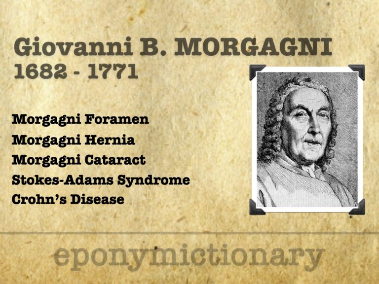
Giovanni Battista Morgagni (1682–1771), father of pathology, pioneered clinico-anatomical correlation; his De sedibus shaped modern medicine.

Vincenz Alexander Bochdalek (1801–1883), Bohemian anatomist who described congenital diaphragmatic hernia and the choroid plexus ‘flower basket’.
Abdominal Aortic Aneurysm (AAA) Surveillance Chart. All incidentally found aortic aneurysms should be referred to a vascular surgeon if the patient is a potential candidate for surgery.
Acute Aortic Dissection (AAD) is uncommon but highly lethal, requiring prompt recognition and management. Due to its non-specific clinical presentation, a high index of suspicion is necessary, particularly in high-risk patients.
Adolphe Pinard (1844–1934) was a French obstetrician. Inventor of the Pinard horn (fetoscope) and Pinard Obstetric Palpation
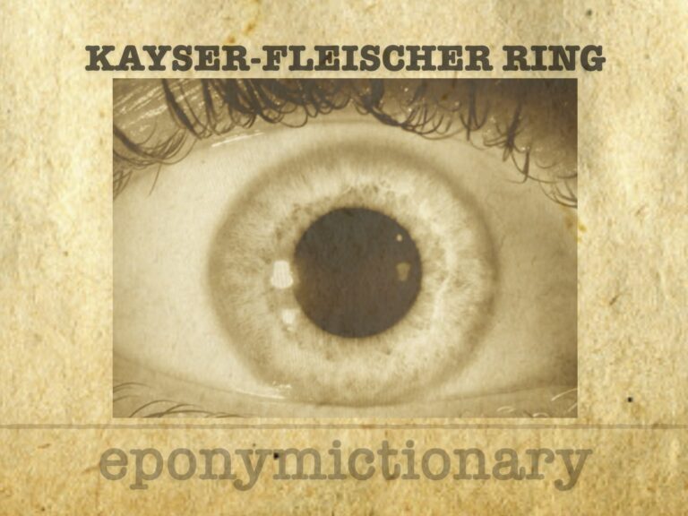
corneal ring at the level of Descemet’s membrane, caused by copper deposition in the cornea. It is a cardinal sign of Wilson’s disease (hepatolenticular degeneration)
The Aortic Dissection Detection Risk Score (ADD-RS) is a validated scoring system that helps stratify low to moderate risk patients who may have an aortic dissection.