
![]()
Chilaiditi syndrome
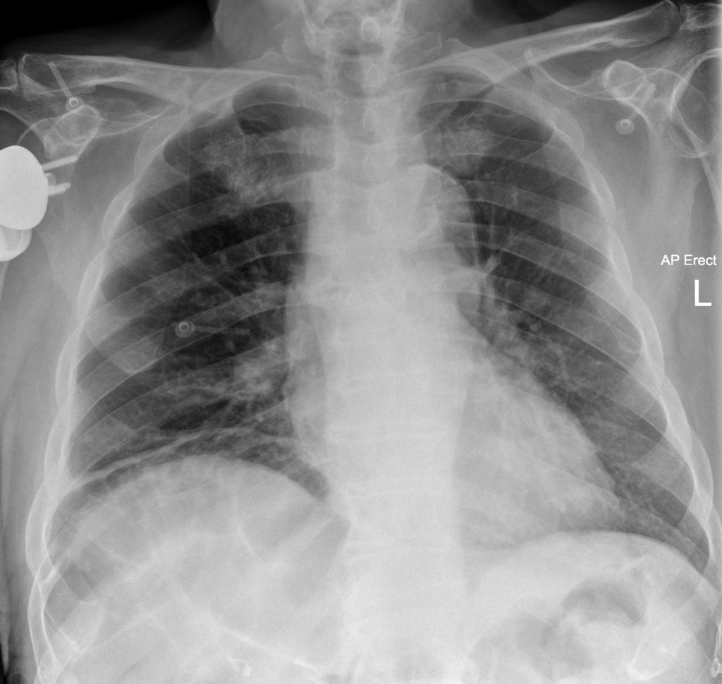
Chilaiditi sign refers to a radiological finding where a loop of colon (or, less commonly, small bowel) is interposed between the liver and right hemidiaphragm, simulating free air on plain radiography. Hepatodiaphragmatic interposition of the intestine (HDI) is typically asymptomatic and discovered incidentally.
When this anatomical anomaly causes symptoms such as abdominal pain, distension, nausea, or vomiting, it is termed Chilaiditi Syndrome.
Clinical Presentation and Epidemiology
Incidence: Chilaiditi’s sign is rare, occurring in 0.1–1% of the general population. The incidence of symptomatic Chilaiditi syndrome is even lower, estimated at 0.025–0.28%.
Demographics: More common in males, elderly, and individuals with chronic constipation, or neurological impairments such as intellectual disability. Associated with liver disease, particularly cirrhosis. Found more frequently in patients with neurological disorders or institutionalised individuals due to altered abdominal muscle tone or posture
Symptoms: Variable and often nonspecific and include nausea, vomiting, abdominal pain, constipation, dysphagia, respiratory distress, and in some cases, signs mimicking acute abdomen.
Aetiology
Chilaiditi’s sign occurs due to anatomic or functional factors that permit colonic interposition between the liver and diaphragm. These include:
- Elongated colon (redundant colon) with long mesentery
- Laxity of suspensory ligaments (e.g., falciform or hepatic ligaments)
- Liver atrophy or reduced liver size — particularly due to cirrhosis
- Chronic constipation and colonic dysmotility
- Chronic obstructive pulmonary disease (COPD) or diaphragmatic elevation
- Neuromuscular conditions, intellectual disability, or spinal deformities
- Previous abdominal surgery (e.g., gastrectomy, hepatic resection)
- Ascites resolution in cirrhotic patients (liver volume decrease)
Patients with cirrhosis and ascites who undergo therapeutic paracentesis are at increased risk of developing Chilaiditi’s sign due to liver shrinkage and colonic mobility
Diagnosis
Radiological Criteria for Chilaiditi’s Sign include:
- Interposition of gas-filled colon between the liver and diaphragm
- Identification of haustral markings, differentiating gas within bowel from pneumoperitoneum
- No change in gas position with patient movement — unlike free air.
Imaging modalities:
- Chest X-ray – classic subdiaphragmatic air with haustral markings
- Abdominal X-ray
- Ultrasound – gas echoes over liver without shifting with position
- CT scan – confirms bowel loop interposition and rules out perforation.
Management
- Asymptomatic (Chilaiditi sign) – No treatment required
- Symptomatic (Chilaiditi syndrome) – Treatment depends on severity:
- Conservative: Bowel rest, fluids, enemas, nasogastric decompression
- Surgical (rare): Colectomy, colopexy, or hemicolectomy if complicated by volvulus or bowel ischaemia.


History of Chilaiditi syndrome
1866 – Arnoldo Cantani (1837-1893) first described the interposition of the intestine between the liver and the diaphragm on clinical examination in his Caso di fegato ambulante. Referenced by Schmidt’s Jahrbücher in 1869 as Fall von wandernder Leber and later by Rogers in 1935.
Il fegato, ridotto di volume, veniva spinto dal sussidio dell’intestino tra il diaframma e la base del torace…
Cantani 1866
“The liver, reduced in volume, was pushed by the intestine between the diaphragm and the base of the thorax…”
Note: This description predates radiographic era (1895) and is interpreted as early recognition of visceral interposition on clinical grounds.
1899 – Antoine Béclère (1856-1939) published the first radiological misdiagnosis of subphrenic gas shadow that autopsy later confirmed to be bowel interposed above the liver. The first published post-mortem confirmation of colonic interposition mimicking gas under the diaphragm.
L’autopsie… a montré qu’une ectopie du côlon transverse venant, à certains moments, s’interposer entre le foie et le diaphragme, était la cause de la singulière image observée à l’examen radioscopique…
Béclère 1899
“The autopsy… revealed that an ectopic transverse colon, at times interposing itself between the liver and the diaphragm, was the cause of the unusual image seen on radioscopic examination…”
1907 – Max Cohn (1880-1915) Presented early radiological insights into colonic displacement and altered hepatic topography in gaseous distension at the third meeting of the meeting of the German Radiological Society
Bei allgemeinem Meteorismus kann der Querdarm so weit nach oben verlagert sein, dass er sich zwischen Leber und Zwerchfell schiebt und zu Fehldiagnosen führen kann…
Cohn 1907
“In general meteorism, the transverse colon may be displaced so far upwards that it slips between the liver and diaphragm and may lead to misdiagnosis…”
Note: In this context “general meteorism” refers to diffuse abdominal distension from excessive intestinal gas. Modern equivalents include generalised bloating or widespread colonic distension, which can shift intra-abdominal anatomy and lead to radiographic misinterpretation. In Cohn’s case he identified gas-filled, distended loops of bowel (specifically the transverse colon) could mimic other pathology (e.g. subphrenic abscess or hepatoptosis) on imaging.
1908 – Maximilian Weinberger (1875-1954) provided the first clear differential diagnosis distinguishing colonic interposition from subphrenic abscess or hepatic atrophy using radiographic methods.
Original
English
Schon 1901 habe ich das instruktive Bild eines subphrenischen Gasabszesses beschrieben… sowie auch Béclère auf die Differentialdiagnose dieses Bildes gegenüber atrophischer Leber hingewiesen, wenn sich zwischen das verkleinerte Organ und das Zwerchfell gashaltige Därme eingeschoben haben – Weinberger 1908
As early as 1901, I described the instructive image of a subphrenic gas abscess… and, like Béclère, pointed out the differential diagnosis of this image compared to an atrophic liver, when gas-filled intestines are interposed between the shrunken organ and the diaphragm – Weinberger 1908

1910 – Dimítrios Chilaiditi (1883-1975) reported the anatomo-radiographic findings of 3 asymptomatic cases (2 male, one female) of subdiaphragmatic air on plain XR as a result of temporary hepatodiaphragmatic interposition of the colon.
Die Kolonschlingen hatten sich in den rechten subphrenischen Raum vorgeschoben und lagen zwischen Leber und Zwerchfell.
Chilaiditi 1910
“The loops of the colon had advanced into the right subphrenic space and were positioned between the liver and the diaphragm.”

Middle: Comparative anatomical positioning of colon, liver, and stomach.
Right: Lateral shift of colonic loops with visible stomach (Magen).
Chilaiditi illustrated the anatomical relationships in three patients with detailed sketches and radiographic correlation establishing the radiological identity of the Chilaiditi sign.
1938 – Wilhelm Behrens used the term Chilaiditi-Symptom in describing symptomatic colonic interposition transitioning from purely radiographic to clinical significance.
1946 – Otto Hubacher published one of the earliest large cohort studies on colonic interposition; continues use of the term Chilaiditi-Symptom. Hubacher’s paper helped formalise the idea of an associated symptom complex, though the term “syndrome” was still not used.
The condition is believed to be congenital in origin… Symptoms may consist of subileus, chronic constipation, or cardiac compression — but diagnosis is rare.
Hubacher 1946
1954 – Baum and Karpati used the term “Chilaiditi‑Syndrom” in German literature, signalling the transition from purely radiographic “sign or symptom” to a recognised clinical syndrome.
1957 – Jackson and Hodson published “Chilaiditi’s syndrome” in the English paediatric literature and defined the distinction between an incidental radiological finding and symptomatic presentations in children.
It is suggested that the syndrome of interposition of the colon in children is a definite entity, easily recognized by a typical clinical and radiological picture, and is of some importance by reason of the symptoms it may produce and the possible complication of intestinal obstruction which may lead to an unnecessary laparotomy if the nature of the condition is unrecognized. It is very probable that more cases would be discovered if the existence of the syndrome were more widely appreciated.
Jackson 1957
Post-1950s Developments in Chilaiditi Sign and Syndrome
Advancements in Imaging and Diagnosis
- From the 2000s, ultrasound and CT imaging became essential tools for differentiating Chilaiditi syndrome from pneumoperitoneum.
- Sato et al. (2000) described hallmark ultrasonographic features, emphasizing positional gas echoes that remain static, unlike free air.
- Saber and Boros (2005) stressed the importance of recognizing Chilaiditi sign to avoid unnecessary surgical intervention such as laparotomy.
Contemporary Clinical Relevance
- Most cases are now managed non-operatively, with interventions like bowel decompression or observation.
- Surgical treatment is reserved for complications such as volvulus, bowel obstruction, or ischaemia.
- Modern reviews underscore the need for heightened awareness in differential diagnosis, especially in emergency and geriatric care settings.
Associated Persons
- Arnoldo Cantani (1837-1893) Italian physician
- Antoine Louis Gustave Béclère (1856-1939) French radiologist
- Max Cohn (1880–1915) German radiologist
- Maximilian Weinberger (1875–1954) Austrian physician
- Dimítrios Chilaiditi (1883-1975)
Alternative names
- Hepatodiaphragmatic interposition of the intestine (HDI)
References
Original articles
- Cantani A. Caso di fegato ambulante. Annali universali di medicina 1866; 62(198): 373-382
- Cantani A. Fall von wandernder Leber. Schmidt’s Jahrbücher der in- und ausländischen gesammten Medicin. 1869; 141: 108
- Gontermann. Ein Fall von Wanderleber. Deutsche medizinische Wochenschrift 1890; 16(46): 1043-1044
- Cohn M. Zur Diagnostik der Dickdarmstenose, zugleich ein Beitrag zur Topographie der Leber bei allgemeinem Meteorismus. Verhandlungen der Deutschen Röntgen-Gesellschaft 1907; 3: 66
- Weinberger M. Weitere Beiträge zur Radiographie der Brustorgane. Medizinische Klinik 1908; 4(1): 584-587
- Béclère A. Rectification d’une erreur de diagnostic; ectopie du colon transverse prise, a l’examen radioscopique, pour un abces gazeux sousphrenique. Bulletins et Mémoires de la Société médicale des hôpitaux de Paris. 1899; 16: 506-507. [PDF]
- Chilaiditi D. Zur Frage der Hepatoptose und Ptose im Allgemeinen im Anschluss an Drei Fälle von Temporärer, Partieller Leberverlagerung. Fortschritte auf dem Gebiete der Röntgenstrahlen. 1910; 16: 173-208
- Behrens W. Ein Beitrag zur Interpositio Colonis (Chilaiditi-Symptom). Helvetica Medica Acta 1938; 5: 110.
- Baum G, Karpati A. Zusammenfassendes über das „Chilaiditi-Syndrom“. Summarizing remarks on the Chilaiditi syndrome. Med Monatsschr. 1954 Apr;8(4):221-3
Review articles
- Rogers JCT. Hepato-diaphragmatic interposition of the colon: Report of a case. Illinois Medical Journal. 1935; 68: 264-268.
- Hubacher O. Interposition of the Colon Between the Liver and Diaphragm (the Chilaiditi Symptom). Schweizerische medizinische Wochenschrift. 1946; 76: 554-559
- Jackson AD, Hodson CJ. Interposition of the colon between liver and diaphragm (Chilaiditi’s syndrome) in children. Arch Dis Child. 1957 Apr;32(162):151-8
- Vessal K, Borhanmanesh F. Hepatodiaphragmatic interposition of the intestine (Chilaiditi’s syndrome). Clin Radiol. 1976 Jan;27(1):113-6.
- Melester T, Burt ME. Chilaiditi’s Syndrome: Report of Three Cases. JAMA. 1985; 254(7): 944-945.
- Hountis P, Chounti M. Chilaiditi’s sign or syndrome? Diagnostic question in two patients with concurrent cardiovascular diseases. Monaldi Arch Chest Dis. 2017 Jul 18;87(2):775.
- Moaven O, Hodin RA. Chilaiditi Syndrome – A Rare Entity with Important Differential Diagnoses. Gastroenterol Hepatol (N Y). 2012; 8(4): 276–278.
- Nakagawa H, Toda N, Taniguchi M, Ibukuro K, Tagawa K. Prevalence and sonographic detection of Chilaiditi’s sign in cirrhotic patients without ascites. AJR Am J Roentgenol. 2006 Dec;187(6):W589-93.
- Sato M et al. Chilaiditi syndrome: sonographic findings. Abdom Imaging. 2000 Jul-Aug;25(4):397-9
- Saber AA, Boros MJ. Chilaiditi’s syndrome: what should every surgeon know? Am Surg. 2005 Mar;71(3):261-3.
eponymictionary
the names behind the name
BA MA (Oxon) MBChB (Edin) FACEM FFSEM. Emergency physician, Sir Charles Gairdner Hospital. Passion for rugby; medical history; medical education; and asynchronous learning #FOAMed evangelist. Co-founder and CTO of Life in the Fast lane | On Call: Principles and Protocol 4e| Eponyms | Books |