
![]()
Echo basics: Machines and Probes
We can do transthoracic echocardiography (TTE) pretty much anywhere. This has resulted in a range of different types of machines to reflect this. Here are the pros and cons of 3 types of machines, how to identify the different types of probes, and what each type of probe is used for.
Full-size, high-end echocardiography machines
Full-size echocardiography machines are great for complex echo techniques, and tend to be found in an outpatient environment. They are stationary.
Pros
- Permanently wire to the archive, which makes it much easier to save our studies.
- They provide really good quality images.
Cons
- Heavy
- Difficult to steer
- Tends to be expensive
Laptop-sized echocardiography machines
These are great for going on the wards, using in your emergency department, or taking them out to a community clinic, because they’re so portable.
Pros
- Moderate cost
- Reasonable functionality
- Improving functionality e.g 3D
Cons
- Tend not to be wired to the archive, so you have to actively remember to save your studies at a later date.
- The image quality not the best.
Handheld echo machines
Handheld echo machines are changing the character of echocardiography. They’re about the size of a mobile phone. Most can do imaging, and some can do colour.
Pros
- Highly portable
- Affordable
Cons
- Limited functionality
- Storage is limited
- Difficult to use, especially for the novice
What are the different types of probes?
Now let’s think about the probes. It could be that your machine just has one probe hanging on it, and that’s easy. You know what to do.
But there could be several, so you need to be able to recognize the probe you’re going to use. Here are 4 types of probes:
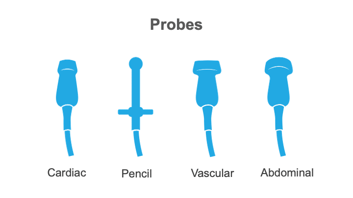
Cardiac ultrasound probes
The bit that you place on the patient’s chest is quite small on a cardiac probe, so it fits between the patient’s ribs. On a paediatric probe, it’ll be even smaller.
Pencil probes
These probes are long and skinny, with a circular end. We use this type of probe for standalone continuous wave Doppler.
Vascular imaging probes
If you see a probe with a long, skinny footprint, it’s probably going to be used for vascular imaging.
Abdominal ultrasound probes
And if you see a probe with a big footprint and a curved shape to it, that’s probably going to be used for abdominal ultrasound.
What frequency is used for different probes?
You need to use the right frequency of probe for what you’re going to use, and for adult practice, that’s going to be between 2 and 4 MHz. For paediatrics, it’ll be higher, at about 7.
This is an edited excerpt from the Medmastery course Echocardiography Essentials by Helen Rimington, PhD. Acknowledgement and attribution to Medmastery for providing course transcripts.
More echocardiography resources:
- Na, M. Echo Masterclass: Left Ventricular Strain. Medmastery
- Monteiro, C. Echo Masterclass: The Right Heart. Medmastery
- Eggett, C. Echo Masterclass: The Valves. Medmastery
- West, C. Echo Masterclass: Adult Congenital Heart Disease. Medmastery
- Naderi, H. Echo Masterclass: The Power of 3D Imaging. Medmastery
Radiology Library: Echocardiography basics
- Rimington H. Echo basics: Machines and probes. LITFL
- Rimington H. Echo basics: Tips and Tricks. LITFL
- Rimington H. Echo basics: Key concepts and Doppler modalities. LITFL
- Rimington H. Echo basics: Parasternal Views. LITFL
- Rimington H. Echo basics: Apical and Subcostal Views. LITFL
Echocardiography Essentials

Helen is a Consultant Cardiac Physiologist at Guy’s and St Thomas’ NHS Trust, in London (UK). She's also a co-author of Echocardiography: A Practical Guide for Reporting. | Medmastery