
![]()
Echo basics: Parasternal Views
Echocardiography Views
Patient position coupled with probe placement and orientation for optimal parasternal long-axis (PLAX) and parasternal short-axis (PSAX) views. See next post for apical and subcostal views
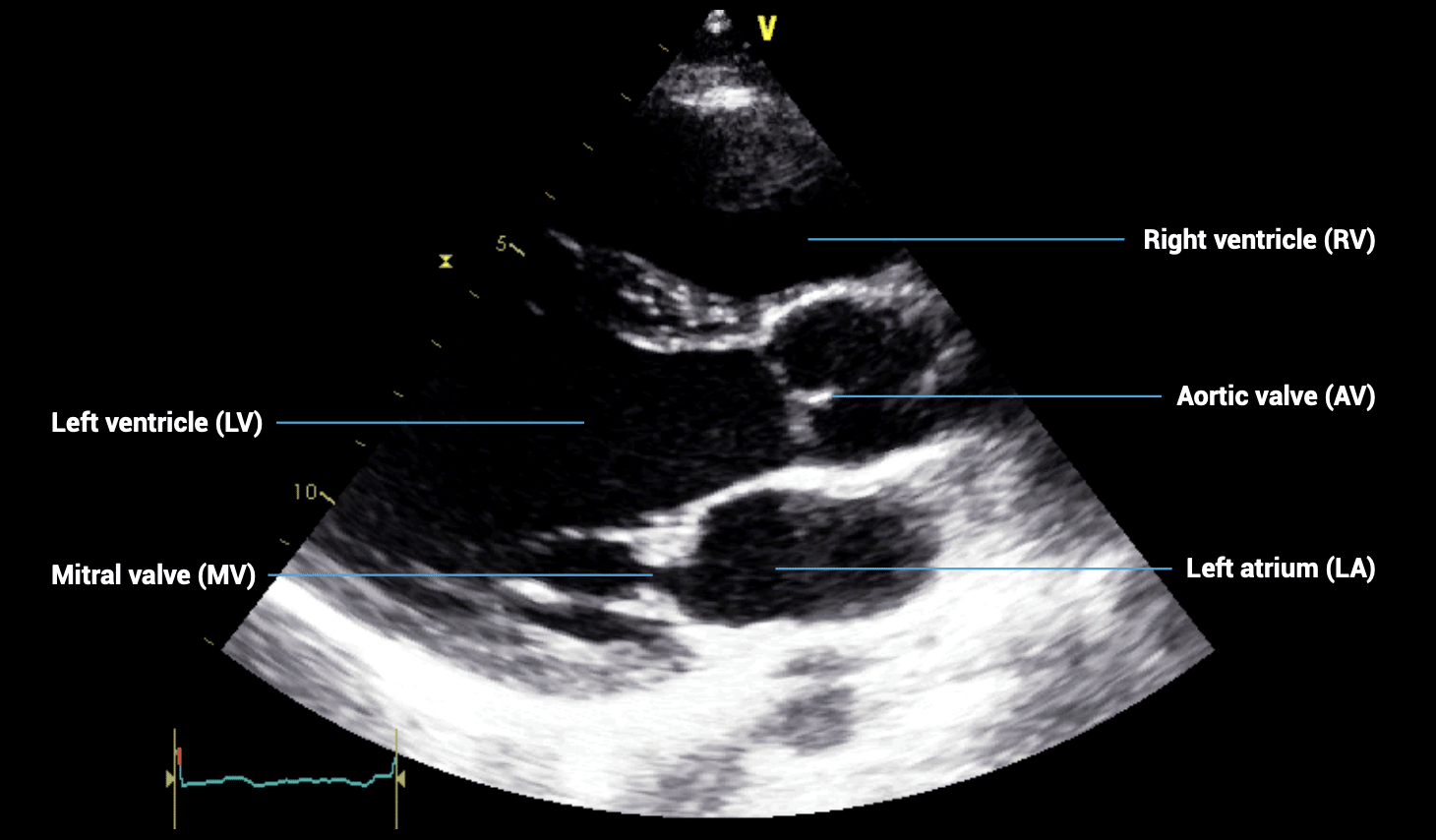
PARASTERNAL LONG-AXIS (PLAX) VIEWS – Obtaining an optimal image
- Patient position: lying on left side, left arm raised, raise the back of the bed or use pillows under the left shoulder
- Probe position: 4th intercostal space, left sternal edge
- Probe orientation: notch towards patient’s right shoulder
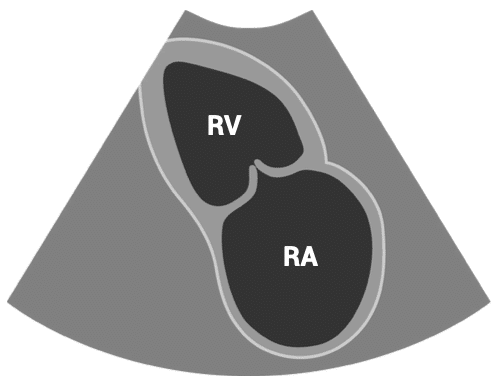
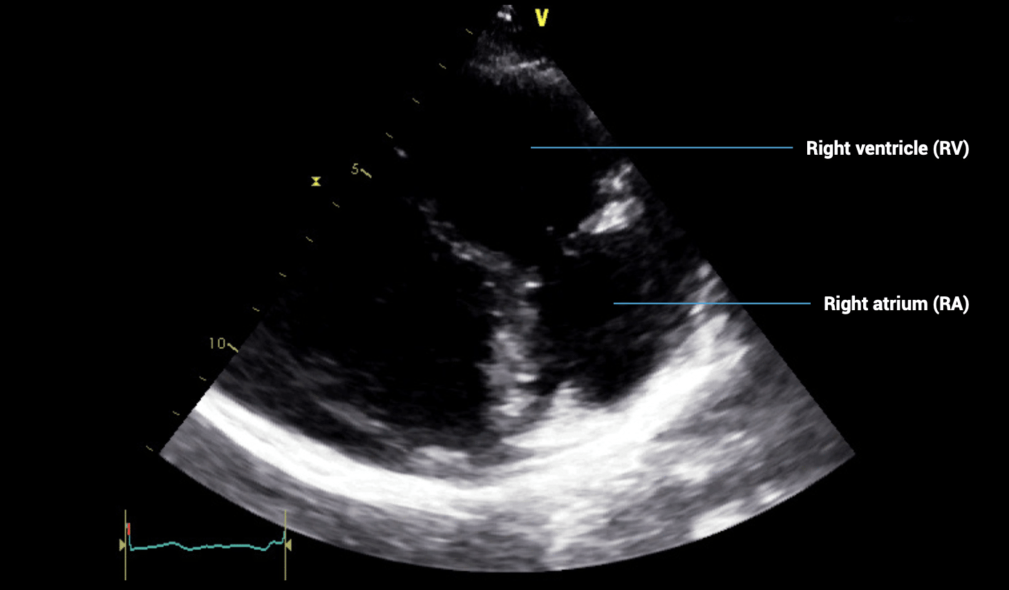
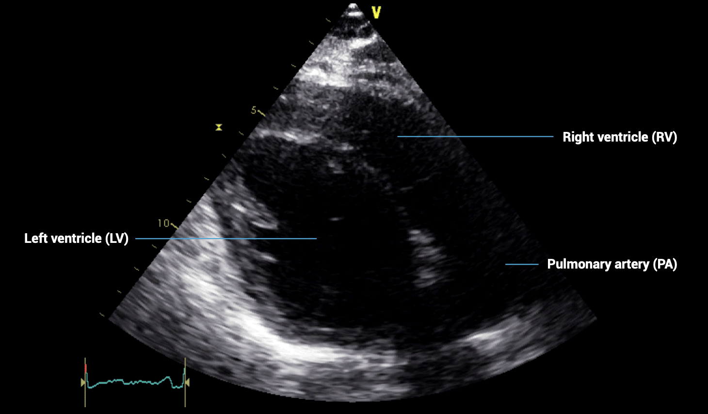
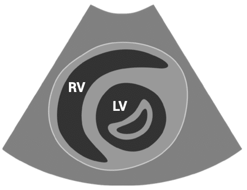
PARASTERNAL LONG-AXIS (PLAX) VIEWS – PLAX inflow view
- Patient position: lying on left side, left arm raised, raise the back of the bed or use pillows under the left shoulder
- Probe position: 4th intercostal space, left sternal edge
- Probe orientation: notch towards patient’s right shoulder
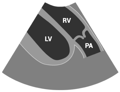
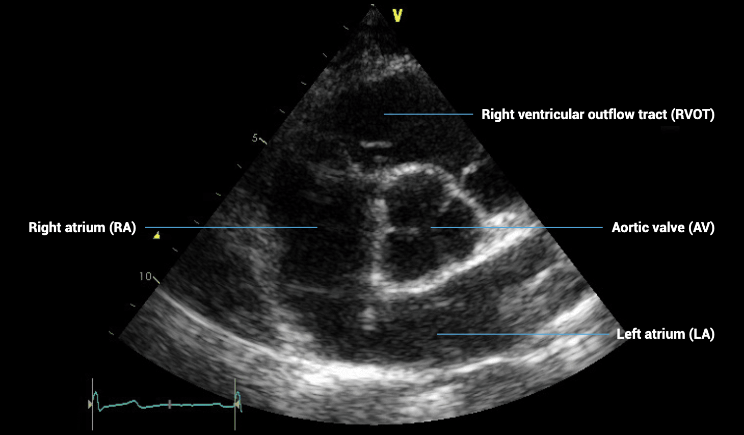
PARASTERNAL LONG-AXIS (PLAX) VIEWS – PLAX outflow view
- Patient position: lying on left side, left arm raised, raise the back of the bed or use pillows under the left shoulder
- Probe position: 4th intercostal space, left sternal edge
- Probe orientation: notch towards patient’s right shoulder
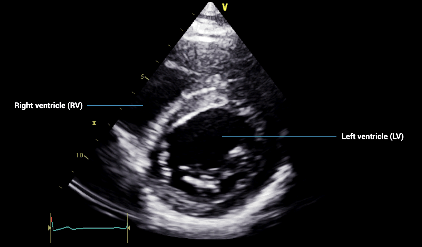
PARASTERNAL SHORT-AXIS (PSAX) VIEWS – PSAX aortic valve view
- Patient position: lying on left side, left arm raised, raise the back of the bed or use pillows under the left shoulder
- Probe position: 4th intercostal space, left sternal edge
- Probe orientation: notch towards patient’s left shoulder
Tilt probe upwards towards head to show aorta as a circle.
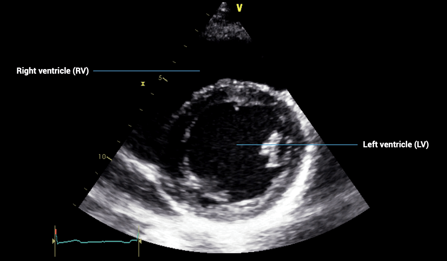
PARASTERNAL SHORT-AXIS (PSAX) VIEWS – PSAX mitral valve view
- Patient position: lying on left side, left arm raised, raise the back of the bed or use pillows under the left shoulder
- Probe position: 4th intercostal space, left sternal edge
- Probe orientation: notch towards patient’s left shoulder
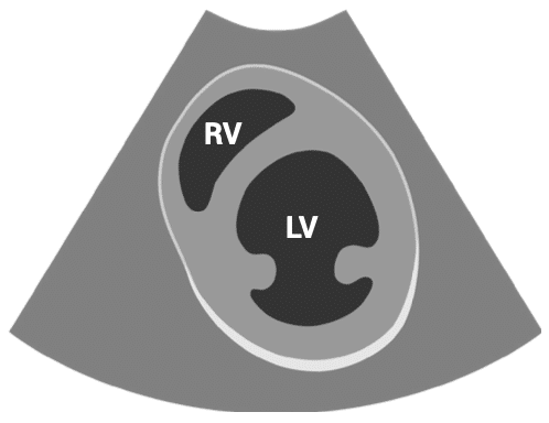
Tilt probe downwards towards spine to show left ventricle as a perfect circle.
PARASTERNAL SHORT-AXIS (PSAX) VIEWS – PSAX papillary muscle view
- Patient position: lying on left side, left arm raised, raise the back of the bed or use pillows under the left shoulder
- Probe position: 4th intercostal space, left sternal edge
- Probe orientation: notch towards patient’s left shoulder
Tilt the probe even further towards the spine keeping the left ventricle as a circle but showing the papillary muscles instead of the mitral valve.
This is an edited excerpt from the Medmastery course Echocardiography Essentials by Helen Rimington, PhD. Acknowledgement and attribution to Medmastery for providing course transcripts.
Additional echocardiography resources:
- Na, M. Echo Masterclass: Left Ventricular Strain. Medmastery
- Monteiro, C. Echo Masterclass: The Right Heart. Medmastery
- Eggett, C. Echo Masterclass: The Valves. Medmastery
- West, C. Echo Masterclass: Adult Congenital Heart Disease. Medmastery
- Naderi, H. Echo Masterclass: The Power of 3D Imaging. Medmastery
Radiology Library: Echocardiography basics
- Rimington H. Echo basics: Machines and probes. LITFL
- Rimington H. Echo basics: Tips and Tricks. LITFL
- Rimington H. Echo basics: Key concepts and Doppler modalities. LITFL
- Rimington H. Echo basics: Parasternal Views. LITFL
- Rimington H. Echo basics: Apical and Subcostal Views. LITFL
Further reading
- European Society of Cardiology. 2017. The EACVI Textbook of Echocardiography 2017 (2e). Lancellotti P, Zamorano JL, Habib G, and Badano L (Eds). Oxford, UK: Oxford University Press.
- Anderson, B. Echocardiography: The Normal Examination and Echocardiographic Measurements 2006 (2e). Hoboken, NJ: Wiley-Blackwell.
- Wharton G, Steeds R et al. A minimum dataset for a standard adult transthoracic echocardiogram: a guideline protocol from the British Society of Echocardiography. Echo Res Pract. 2015 Mar 1;2(1):G9-G24.
Echocardiography Essentials

Helen is a Consultant Cardiac Physiologist at Guy’s and St Thomas’ NHS Trust, in London (UK). She's also a co-author of Echocardiography: A Practical Guide for Reporting. | Medmastery