![]()
Hoffman-Rigler sign
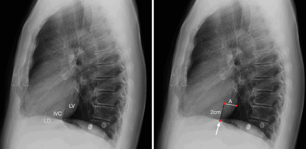
A radiological sign used on the lateral chest X-ray to detect left ventricular enlargement (LVE). The sign is based on anatomical landmarks involving the left ventricle (LV) and inferior vena cava (IVC):
- A vertical line is drawn 2 cm cephalad from the intersection of the LV and IVC on the lateral projection.
- A second line is drawn posteriorly, parallel to the vertebral bodies.
- If the posterior border of the LV extends more than 1.8 cm beyond the IVC at this level, LVE is suggested.
Note: Eyler et al described the method in 1959, but Hoffman and Rigler standardized and popularized the measurement. There is no evidence of plagiarism, more a case of refinement and validation.
History of the Hoffman-Rigler sign
1959 – Eyler et al first proposed that posterior LV extension beyond the IVC in lateral view suggests enlargement, especially in rheumatic heart disease.
In patients with rheumatic lesions of the mitral valve, the detection of left ventricular enlargement in the lateral view has been the most reliable single roentgen indication of the presence of mitral insufficiency or of an aortic valve lesion. All four views as well as fluoroscopy should be used.
When the left ventricle was enlarged, it has in most cases projected behind the shadow of the inferior vena cava for a distance of 15 mm or more.
Eyler et al, Radiology 1959
1965 – Richard Bashefkin Hoffman (1937-2011) and Leo George Rigler (1896-1979) published their paper defining two measurements easily obtained from the lateral chest film and to determine their degree of efficacy in evaluating left ventricular size.
Measurement A is defined as the distance which the left ventricle extends posteriorly to the posterior border of the inferior vena cava at a point 2 em cephalad to the crossing of the cava and the left ventricle. Measurement is made on a plane extending posteriorly which parallels the horizontal plane of the vertebral bodies.
Measurement B is the distance of the crossing, referred to above, caudad to the left leaf of the diaphragm
When the posterior border of the left ventricle extends posteriorly to the posterior border of the inferior vena cava more than 1.8cm at a level 2cm cephalad to their crossing, on a lateral projection of the chest in the adult, one can postulate left ventricular enlargement with a considerable degree of certainty
Hoffman, Rigler Radiology 1965

Fig. 2. Right lateral projection of a normal heart.
A. Radiograph with dots designating limits of measurements A and B.
B. Diagram of the posterior inferior cardiac border of the same case with measurements A and B designated.
LV = left ventricle; IVC = inferior vena cava; RD = right diaphragm; LD = left diaphragm

B. Right lateral view demonstrates large measurement
C. Diagram illustrating measurements
Validation and Modern Use
2008 – Serna et al evaluated the validity of the H&R sign against the echocardiographic LV end-diastolic diameter in 200 patients and showed a sensitivity of 92.5%, a specificity of 83.3%, a positive predictive value of 78.7%, and a negative predictive value of 94.3%.
They conclude that the Hoffman-Rigler sign is a valuable alternative in the evaluation of left ventricular enlargement when cardiac ultrasound is not readily available
2016 – Spaziano et al investigated the sensitivity and specificity of the Hoffman-Rigler sign in a modern population. A sample of 145 patients with LV dilatation was matched for age and sex with 145
patients without LV dilatation. The H&R sign and the cardiothoracic index were assessed on the radiograph independently by 2 blinded physicians.
Spaziano et al found that while measurable in two-thirds of patients, the sign showed low diagnostic accuracy in modern populations.
Associated Persons
- Leo George Rigler (1896-1979)
- Richard Bashefkin Hoffman (1937-2011)
References
Historical references
- Eyler WR, Wayne DL, Rhodenbaugh JE. The importance of the lateral view in the evaluation of left ventricular enlargement in rheumatic heart disease. Radiology. 1959 Jul;73(1): 56-61.
- Keats TE, Rudhe U, Foo GW. Inferior vena caval position in the differential diagnosis of atrial and ventricular septal defects. Radiology. 1964 Oct;83: 616-621.
- Hoffman R, Rigler L. Evaluation of the left ventricular enlargement in the lateral projection of the chest. Radiology 1965: 85: 93–100.
Eponymous term review
- Freeman V, Mutatiri C, Pretorius M, Doubell A. Evaluation of left ventricular enlargement in the lateral position of the chest using the Hoffman and Rigler sign. Cardiovasc J S Afr. 2003 May-Jun;14(3):134-7
- Serna GG, Villarosa AF. Validity of Hoffman and Rigler sign in the evaluation of left ventricular enlargement. Phil Heart Center J 2008;14: 39-41
- Spaziano M, Marquis-Gravel G, Ramsay I, Romanelli G, Marchand É, Tournoux F. Left Ventricular Dilatation Assessed on the Lateral Chest Radiograph: The Classic Hoffman and Rigler Sign Falls Short in a Modern-Day Population. Can J Cardiol. 2016 Mar;32(3):378-83
eponymictionary
the names behind the name
BA MA (Oxon) MBChB (Edin) FACEM FFSEM. Emergency physician, Sir Charles Gairdner Hospital. Passion for rugby; medical history; medical education; and asynchronous learning #FOAMed evangelist. Co-founder and CTO of Life in the Fast lane | On Call: Principles and Protocol 4e| Eponyms | Books |