
![]()
John Howship
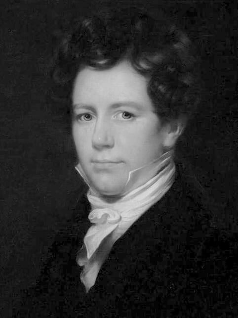
John Howship (1781-1841) was an English surgeon.
Howship was a surgeon and anatomist whose work advanced surgical pathology and systemic clinical observation in the early 19th century. Trained under John Heaviside, he developed a strong foundation in dissection and morbid anatomy, which shaped his approach to diagnosis and surgical practice.
Howship contributed seminal observations in multiple domains: he first documented the phenomenon later recognized as a growing skull fracture, described bone resorption lacunae that bear his name, and introduced diagnostic principles for gastrointestinal and urinary disorders. His writings such as Practical Observations on Diseases of the Urinary Organs (1816) and Practical Observations on the Diseases of the Lower Intestines and Anus (1820) provided systematic guidance at a time when surgical specialization was in its infancy.
He is most widely remembered today for the Howship–Romberg sign, an important diagnostic clue for obturator hernia, and for his contributions to understanding bone physiology. Howship’s career also reflects the turbulent institutional politics of early Victorian medicine, culminating in his appointment as Chief Surgeon at Charing Cross Hospital. He died suddenly in 1841, leaving a legacy embedded in surgical practice and clinical signs that endure in modern textbooks.
Biographical Timeline
- 1781 – Born in London, England.
- 1799 – Entered medical school; apprenticed under John Heaviside (1748–1828), focusing on anatomical dissection and specimen preparation. Practised ‘Museum Medicine’ honing dissection skills to define anatomical sites of disease
- 1805 – Appointed Assistant Surgeon at St George’s Infirmary, London; began lecturing at St George’s Hospital School.
- 1815 – Published early studies on bone formation and union.
- 1816–1825 Issued major works on urinary diseases, gastrointestinal pathology, and bone disorders, including:
- 1816 – Published Practical Observations in Surgery and Morbid Anatomy, describing partial absorption of the parietal bone in infants after trauma—the first account of what is now termed growing skull fracture (GSF).
- 1828 – Elected to the Council of the Royal College of Surgeons; became prominent in London surgical circles.
- 1833 – Delivered the Hunterian Lecture at the Royal College of Surgeons; published an illustrated surgical atlas.
- 1834–1836 Appointed Assistant Surgeon (1834) then Chief Surgeon (1836) at Charing Cross Hospital, following the dismissal of Thomas Pettigrew.
- 1840 – Published Practical Remarks on the Discrimination and Appearance of Surgical Disease, including observations later linked to the Howship–Romberg sign.
- Died January 22, 1841 aged 59, from haemorrhage secondary to abscess of the tibia.
Medical Eponyms
Howship-Romberg sign (1840, 1847)
Pain and paraesthesia along the distribution of the obturator nerve, classically radiating from the inner (medial) aspect of the thigh, down to the knee. Pain is exacerbated by thigh extension, abduction, and internal rotation of the hip, and relieved by thigh flexion. It is a clinical indicator of obturator nerve compression, most frequently due to an obturator hernia, but can rarely result from other pelvic masses such as inflammatory granuloma.
Obturator hernia is rare, accounting for less than 1% of all abdominal hernias, and is most commonly seen in elderly, thin, multiparous women. The sign is present in 30–50% of cases and should raise suspicion in patients with symptoms of small-bowel obstruction without obvious cause.
Pathophysiologically, the sign arises from entrapment or irritation of the obturator nerve within the obturator canal by the hernia sac (or other lesion). The pain may mimic hip or knee arthritis, contributing to delayed diagnosis.
1840 – Howship first described the clinical phenomenon in an 1840 case report titled “Strangulated Thyroidal Hernia – Diagnostic Symptom – Appearances on Dissection”. He describes then case of an aged and emaciated female under the care of Mr. Weatherfield
…she was seized with violent spasmodic pain in the left side of the abdomen running down the left leg, with sickness, vomiting, and diarrhoea…The symptoms, those of strangulated hernia…She was bled, and directed various aperients and purgatives, a blister and leeches to the abdomen but towards the close of four days of severe suffering, sunk and died.
Howship 1840: 323
Postmortem examination revealed:
…a portion of small intestine was seen stretched towards the obturator foramen, where a knuckle was firmly impacted, forming a small hernia, no larger than a nutmeg, protruding through the opening. The intestine, highly inflamed, was almost gangrenous. The parts were carefully removed and admirably dissected; demonstrating the hernia to the best advantage
Howship 1840: 324
Moritz Heinrich Romberg (1795-1873) described his findings of an obturator hernia and their clinical significance in 1847, published in 1848 as ‘Die Operation des eingeklemmten Bruches des eirunden Loches‘ emphasising the value of the nerve symptom in diagnosis
Original
English
Was diesem Falle ein besonderes Interesse verleiht, ist nicht seine Seltenheit, sondern die Aufklärung der Diagnose durch ein Nervenphänomen… Druck und Zerrung des Nervus obturatorius… manifest [durch] Schmerz an der inneren Seite des Oberschenkels… und Unfähigkeit, den Schenkel anzuziehen
Für die Hernia obturatoria ist ein diagnostisches Kriterium, wie es der Verein der Schmerzen und der gestörten Beweglichkeit des Oberschenkels mit der unterbrochenen Permeabilität des Darmcanals darbietet, um so bedeutungsvoller, weil dieser Bruch nur allmälig, gleichsam chronisch entsteht, selten eine von aussen sichtbare Geschwulst bildet, und, wie kein anderer Bruch, temporären, wiederholten Incarcerationen ausgesetzt ist.
What gives this case a special interest is not that it is rare, but the elucidation of the diagnosis by a neurologic phenomenon. Pressure and distortion of the obturator nerve… will manifest [through] pain on the inner side of the thigh… and inability to adduct the thigh
Obturator hernia is associated with pain; disturbed movement of the thigh; and altered intestinal movement. Most important, because this condition is chronic it rarely forms a tumour visible from the outside, and is subjected to temporary, repeated incarceration.
Howship lacunae
Small pits, grooves or depressions or bone pit (>100 microns in length) with an accumulation of osteoclast cells indicative of bone resorption
Howship’s investigations into bone resorption identified depressions in the bony matrix caused by osteoclastic activity, later termed Howship’s lacunae. These findings helped elucidate mechanisms of bone remodelling and pathological osteolysis.
Key Medical Contributions
Growing Skull Fracture (1816)
Howship’s Practical observations in surgery, and morbid anatomy (1816) contains the first recorded description of a growing skull fracture (GSF) a rare paediatric complication of head trauma. He documented Case 10, a 9-month-old infant who fell down stairs, sustaining a 3 × 1-inch depressed fracture of the parietal bone. Initially unconscious, the child received standard treatment of the era: leeches and saturnine lotion (a lead acetate solution) to reduce inflammation.
Two weeks later, the mother noticed pulsations of the brain through the defect, which persisted and enlarged. By age 4, the cranial gap remained undiminished, pulsations were evident on crying or coughing, and left leg weakness persisted, features now recognized as classic for GSF. Howship concluded the progressive enlargement was linked to impaired blood supply and absorption of bone, not simple mechanical pressure:
The aperture in the parietal bone gradually enlarged; its edges softened and became absorbed.
Howship. Partial Absorption of the Parietal Bone, arising from a Blow on the Head. 1816
This condition later acquired many names including traumatic cephalhydrocele, leptomeningeal cyst, andtraumatic meningocele before Pia and Tönnis (1953) standardized the term growing skull fracture. Howship’s original observation remains the cornerstone for understanding this entity, which occurs almost exclusively in children under 3 years and requires prompt surgical repair to prevent neurological sequelae.
Gastrointestinal and Urinary Contributions
Howship authored influential treatises on anorectal and urinary disease. His 1816 monograph addressed urinary obstruction and calculus management, while his 1820 work on rectal disorders advanced operative approaches to fistula, heamorrhoids, and strictures—issues central to 19th-century surgical practice.
Professional Life and Legacy
A prominent figure in London surgery, Howship lectured widely and served on the Council of the Royal College of Surgeons, later becoming Chief Surgeon at Charing Cross Hospital (1836). His illustrated atlases and surgical manuals reinforced anatomy as the foundation for operative precision. Despite lacking a dedicated obituary, Howship’s legacy survives in clinical signs, eponyms, and enduring surgical concepts.
Major Publications
- Howship J. Cases of Tetatnus, with Observations upon the Disease; in Continuation: A Case of LOCK-JAW, from the Irritation of an extensive Wound, in which Opium certainly checked the Progress of the Disease, and probably saved the Life of the Patient. Med Phys J. 1809 Dec;22(130):479-486.
- Howship J. Experiments and observations in order to ascertain the means employed in the animal economy, in the formation of bone. 1815
- Howship J. Practical observations in surgery, and morbid anatomy: illustrated by cases with dissections and engravings. 1816
- Howship J. Observations on Hernia, Illustrated by a Case and Dissection. Med Chir J Rev. 1817 Mar;3(15):177-179.
- Howship J. Practical observations on the symptoms, discrimination, and treatment of some of the most common diseases of the lower intestines, and anus. 1820 [2e 1825]
- Howship J. Practical remarks upon indigestion. 1825
- Howship J. Practical remarks on the discrimination and successful treatment of spasmodic stricture in the colon. 1830
- Howship J. The Hunterian oration; Royal College of Surgeons in London. 1833
- Howship J. Some account of two cases of inflammatory tumour, produced by the deposit of the larva of a large fly (Oestrus humanus) beneath the cutis, in the human subject. 1833
- Howship J. Practical remarks on the discrimination and appearance of surgical disease; with an appendix containing the descriptive catalogue of the author’s collection in pathological anatomy and the Hunterian Oration for 1833. Churchill, 1840
- Howship J. Strangulated Thyroideal Hernia – Diagnostic Symptom – Appearances on Dissection. In: Practical Remarks on the Discrimination and Appearance of Surgical Disease. 1840: 323-4
References
Biography
- Bir SC, Kalakoti P, Notarianni C, Nanda A. John Howship (1781-1841) and growing skull fracture: historical perspective. J Neurosurg Pediatr. 2015; 16(4): 472-6
Eponymous Terms
- Pia HW, Tonnis W. Die wachsende Schädelfraktur des Kindesalters [Growing skull fractures of childhood]. Zentralbl Neurochir. 1953;13(1):1-23.
- Rizk TA, Deshmukh N. Obturator hernia: a difficult diagnosis. South Med J. 1990; 83(6): 709-12.
- Yamashita K, Hayashi J, Tsunoda T. Howship-Romberg sign caused by an obturator granuloma. Am J Surg. 2004; 187(6): 775-6.
- Domenico Bertoloni Mel. Visualizing Disease. The Art and History of Pathological Illustrations. University of Chicago Press, 2017: 160-162
- Rastogi V et al. Abdominal Physical Signs and Medical Eponyms: Movements and Compression. Clin Med Res. 2018; 16(3-4): 76-82.
Eponym
the person behind the name
BA MA (Oxon) MBChB (Edin) FACEM FFSEM. Emergency physician, Sir Charles Gairdner Hospital. Passion for rugby; medical history; medical education; and asynchronous learning #FOAMed evangelist. Co-founder and CTO of Life in the Fast lane | On Call: Principles and Protocol 4e| Eponyms | Books |