![]()
POCUS Made Easy: Lung
Lung Ultrasound Examination looks for pulmonary pathology including acute pulmonary oedema, pneumonia, pleural effusion, pleural abnormalities such as pneumothorax
Pre-reading
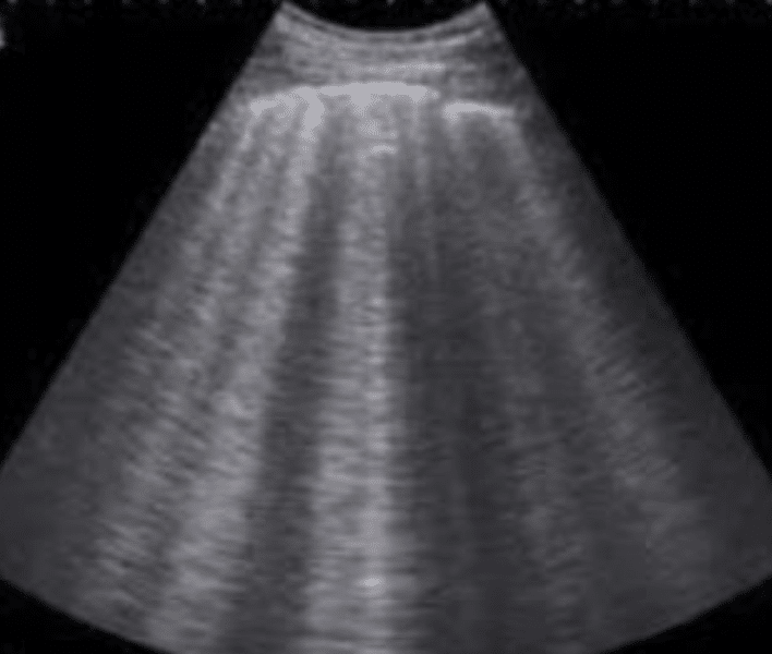
1. Rt Anterior Superior Chest
- Labelled RIGHT ANT SUP
- Longitudinal view
- Demonstration of pleural sliding
- Demonstration of lung field (most sensitive for pneumothorax assessment)
2. Rt Anterior Inferior Chest
- Labelled RIGHT ANT INF
3. Rt Lateral Superior Chest • Labelled RIGHT LAT SUP
4. Rt Lateral Inferior Chest
- Labelled RIGHT LAT INF
- + assess lung-diaphragm interface (more sensitive for dependent lung pathology)
5 to 8. Same as Above on Left Chest *LEFT

Image Sets
- Minimum 10 Video Loops
- 8x Lung Field (4 each side)
- 2x Pleural Assessment (1 each side)
- 10-14 Video Loops +/- 2 Images
- Optional extra views including:
- Posterior Inferior and Superior Views
- Focussed views to highlight Pathology
Machine Settings
- CURVILINEAR Probe
- LUNG setting for all images
Documentation – POCUS LUNG
- Views: Adequate/ Inadequate
- Findings: NAD / Abnormal
- Pleura: NAD / Abnormal (no pleural sliding / lung point / barcode sign on M-Mode / thickened/ irregular)
- Lung Fields: NAD / Abnormal (B-Lines / Effusion / Hepatisation / Shred Sign / Other – and describe)
Anatomical views


Diagnostic Flow Chart

Positive Findings (always consider clinical context)


References and Further reading
- Rippey J. Lung ultrasound Modules. Ultrasound Library
- Rippey J. Lung Clinical Cases. Ultrasound Library
- Bendigo ED Resource PDF – Lung
[cite]

POCUS
made easy

MBChB FACEM CCPU. Emergency Physician at Bendigo Hospital and Royal Melbourne Hospital, Victoria. Australia.
I studied in Scotland before completing my Emergency Medicine training in Melbourne. I have a big interest in point-of-care ultrasound use and training in Emergency Medicine. I'm also interested in Choosing Wisely and sustainability initiatives in healthcare.
In my free time you'll find me playing tennis and soccer, or heading outdoors on camping trips.