
![]()
Risky Rhythyms
aka ECG Exigency 003
5 risky rhythms. Each tells a story. Can you work out what is happening before its too late? What can you do to save the day?
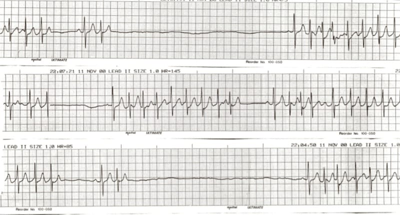
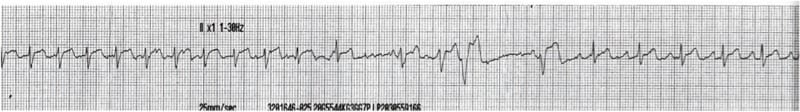
Rhythm strip 1
Q1.1 What rhythm is shown?
This is a classic example of sick sinus with the “tachy-brady” syndrome
- Runs of tachycardia interspersed with long sinus pauses (up to 6 seconds).
- The sinus rate is extremely slow, varying from 40 bpm down to around 10 bpm in places.
- Sinus beats are followed by paroxysms of junctional tachycardia at around 140 bpm.
Q 1.2 What will you do next?
This patient needs a pacemaker, stat!
- Admit to a monitored bed on a coronary care unit.
- Commence temporary pacing via external pads or pacing wire until a permanent pacemaker can be arranged.
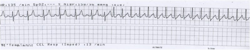
Rhythm strip 2
Q 2.1 What rhythm is shown?
- Six beats of sinus rhythm at 90 bpm.
- The 7th beat is a premature atrial complex (PAC) with different morphology P, QRS and T waves, which initiates a run of a supraventricular tachycardia at 150bpm.
- The onset of the SVT is typical of an AV-nodal re-entry tachycardia (AVNRT), although with the rate of 150bpm, atrial flutter with a 2:1 block is also a possibility.
Q 2.2 What will you do next?
- Scrutinise the 12-lead ECG for flutter waves.
- Try some adenosine (or vagal stimuli such as a Valsalva manoeuvre or carotid massage); this should unmask any flutter waves and may convert AVNRT to sinus rhythm.
- Flutter may require DC cardioversion or treatment with anti-arrhythmics (e.g. amiodarone).
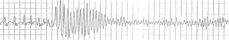
Rhythm Strip 3
Q 3.1 What rhythm is shown?
A narrow complex tachycardia is interrupted by a run of polymorphic VT, which rapidly deteriorates into ventricular fibrillation.
Q 3.2 What will you do next?
- Precordial thump!
- Start charging the defibrillator!
- Shock at 200 J (biphasic) or 360 J (monophasic). Three stacked shocks if the arrest is witnessed and monitored.
- Start CPR!
Rhythm Strip 4
Q 4.1 What rhythm is shown?
- Sinus rhythm, or possibly ectopic atrial rhythm (biphasic / inverted P waves in lead II)
- Rate of 90 bpm
- Prolonged QTc interval of 540 ms (greater than half the R-R interval)
- Ventricular ectopics with ‘R-on-T’ phenomenon
- The second ventricular ectopic initiates a run of torsades de pointes
Q 4.2 What will you do next?
- DC cardioversion if unstable.
- Load with magnesium (e.g. 2 g over 1-2 minutes) and start a magnesium infusion.
- Correct hypokalemia.
- Consider: Overdrive pacing to achieve a ventricular rate of 90-120 bpm.
- Consider: Isoprenaline infusion.
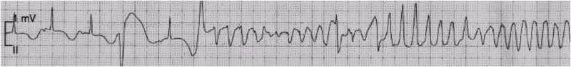
Rhythm Strip 5
Q 5.1 What rhythm is shown?
This is a typical ECG recording of a patient receiving a bolus of adenosine for AVNRT.
- AV-nodal re-entry tachycardia (AVNRT) at 140 bpm
- A pause in the middle of the strip with several ventricular escape complexes
- Cardioversion to sinus rhythm at 90 bpm at the end of the strip
Q 5.2 What will you do next?
- Get a 12-lead ECG to confirm return to sinus rhythm.
- If the patient is well and remains in sinus rhythm they can be discharged.
- Consider electrophysiology follow up for recurrent AVNRT.
References
Further Reading
- Kosuge M, Kimura K, Ishikawa T, Ebina T, Hibi K, Kusama I, Nakachi T, Endo M, Komura N, Umemura S. Electrocardiographic differentiation between acute pulmonary embolism and acute coronary syndromes on the basis of negative T waves. Am J Cardiol. 2007 Mar 15;99(6):817-21.
Further Reading
- Wiesbauer F, Kühn P. ECG Mastery: Yellow Belt online course. Understand ECG basics. Medmastery
- Wiesbauer F, Kühn P. ECG Mastery: Blue Belt online course: Become an ECG expert. Medmastery
- Kühn P, Houghton A. ECG Mastery: Black Belt Workshop. Advanced ECG interpretation. Medmastery
- Rawshani A. Clinical ECG Interpretation ECG Waves
- Smith SW. Dr Smith’s ECG blog.
- Wiesbauer F. Little Black Book of ECG Secrets. Medmastery PDF
CLINICAL CASES
ECG EXIGENCY
CLINICAL CASES
ECG EXIGENCY
Emergency Physician in Prehospital and Retrieval Medicine in Sydney, Australia. He has a passion for ECG interpretation and medical education | ECG Library |