
![]()
Adriaan van den Spiegel

Adriaan van den Spiegel (1578-1625) was a Flemish anatomist and botanist.
Spiegel’s anatomical studies were published posthumously in De humani corporis fabrica libri decem (1627), edited by Daniel Bucretius and illustrated with the plates of Giulio Casseri (1552–1616). This atlas preserved his detailed accounts of the abdominal wall, liver, and viscera. Among his lasting contributions are the descriptions of the Spigelian line and Spigelian fascia, where Spigelian hernias may develop, and the Spigelian lobe of the liver, later termed the caudate lobe (Couinaud’s Segment I).
Spiegel’s influence extended beyond anatomy. The plant genus Spigelia (family Loganiaceae) was named in his honour, and his name remains attached to rare hernias and hepatic anatomy. Despite dying prematurely at 47, his combination of careful description and Casserius’ engravings gave his work an enduring authority.
Biographical Timeline
- 1578 – Born in Brussels, Flemish Brabant.
- Before 1600 – Studied medicine at Louvain and Leiden.
- 1601–1604 – Registered at Padua (Natio Germanica), studied anatomy with Hieronymus Fabricius ab Aquapendente (1533–1619) and Giulio Casseri (1552–1616).
- 1606 – Appointed physician to the German-Dutch student nation at Padua; wrote Isagoges in rem herbariam libri duo, an introduction to botany.
- 1607 – Candidate for chair of practical medicine at Padua (unsuccessful).
- 1612 – Briefly returned to Belgium, then travelled through Germany, settling in Moravia; became medicus primarius of Bohemia.
- 1616 – Appointed Professor of Anatomy and Surgery at Padua by the Venetian Senate.
- 1623 – Elected Knight of the Venetian order of Saint Marco.
- 1624 – Published De semitertiana libri quatuor, first accurate description of malaria.
- Died April 7, 1625 in Padua after six weeks of illness; autopsy found intra-abdominal abscess near the caudate lobe.
- 1627 – Posthumous publication of De humani corporis fabrica libri decem, illustrated with Casserius’ plates, edited by Liberali Crema and Daniel Bucretius.
Medical Eponyms
Spigelian Line (linea semilunaris) and Spigelian Fascia
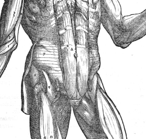
The Spigelian line (linea semilunaris) marks the lateral border of the rectus abdominis, where it is distinguished from the oblique abdominal muscles. Spiegel first described the anatomy, based on the plates of his predecessor Giulio Casseri (1552–1616), in Liber III, Caput IV–VI of De humani corporis fabrica libri decem (1627).
Rectus … ab obliquis utrinque linea quadam semilunari discernitur.
The rectus [muscle] … is separated on each side from the obliques by a certain semilunar line
Spigelii, 1627
The term “linea Spigelii” was applied by anatomical texts in the late 17th–18th century by authors such as Govard Bidloo and William Cheselden.
The Spigelian fascia is the aponeurotic zone formed by the fusion of the internal oblique and transversus abdominis aponeuroses, adjacent to the semilunar line. This area represents a potential weak point in the abdominal wall, predisposing to the Spigelian hernia.
Spigelian Hernia (1764)
A Spigelian hernia is protrusion of peritoneum/abdominal contents through a defect in the Spigelian fascia (between the internal oblique and transversus abdominis aponeuroses) along the linea semilunaris. Most defects occur near or below the arcuate line within the so-called Spigelian belt (6 cm band above the interspinous line), where the posterior rectus sheath thins and creates a physiologic weak zone.
Spigelian hernias are rare (0.12–2% of abdominal wall hernias) predominantly women >60 years. Risk factors include obesity, COPD, pregnancy, multiple pregnancies, and collagen disorders. Incarceration risk is high (17–27%), so even asymptomatic cases are generally scheduled for elective repair to avoid strangulation.
Patients present with localised abdominal wall pain, intermittent swelling, or bowel obstruction. Many patients lack a palpable mass because the intact external oblique can mask the sac.
Diagnosis is challenging. Consider the diagnosis with focal lower abdominal pain, a subtle lateral wall bulge, or obstructive symptoms. Examine standing and with Valsalva although up to 50% are missed on physical exam alone. CT (with oral contrast) is the most reliable test. Ultrasound (standing/Valsalva) is a useful option. Diagnostic laparoscopy is reserved for ambiguous cases.
Many patients lack a palpable mass because the intact external oblique can mask the sac. Consider the diagnosis with focal lower abdominal pain, a subtle lateral wall bulge, or obstructive symptoms; examine standing and with Valsalva. Up to 50% are not confirmed by exam alone.
Most Spigelian hernias are reported to lie in the vicinity of the semicircular line (Fig. 1) and the hernia usually penetrates both the transversus abdominis and internal oblique muscles (Fig. 2).
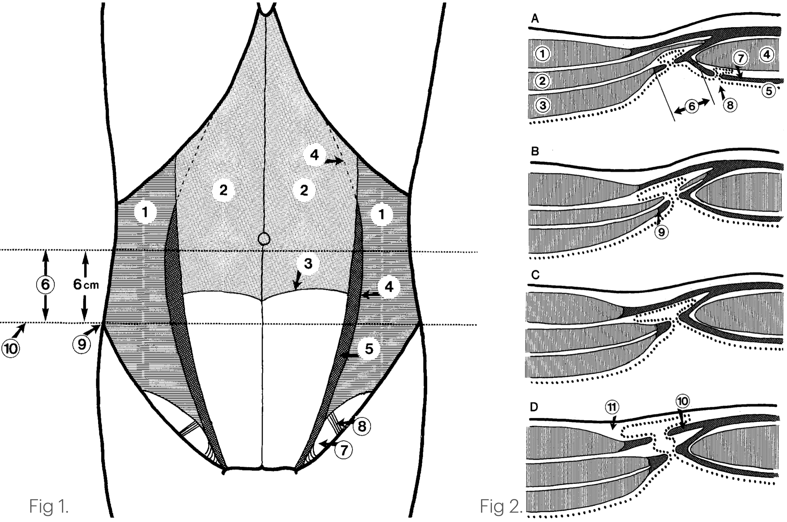
Fig. 1. A ventral view of the abdominal wall showing the topographic anatomy. The external and internal oblique and rectus abdominis muscles are cut away. 1: Transversus abdominis muscle, 2: Dorsal lamella of the rectus sheath, 3: The semicircular line (of Douglas), 4: The semilunar line (Spigelii), 5: Spigelian aponeurosis, 6: Spigelian hernia belt, 7: Hesselbach triangle, 8: Inferior epigastric vessels, 9: Anterior superior iliac spine, 10: Interspinal plane.
Fig. 2. Schematic cross-section of ventral abdominal wall cranial to the semicircular line, indicating the possible location of the hernial sac in spigelian hernias. 1: External oblique muscle, 2: Internal oblique muscle, 3: Transversus abdominis muscle, 4: Rectus abdominis muscle, 5: Peritoneum, 6: Spigelian aponeurosis, 7: Dorsal lamella of the rectus sheath, 8: Intravaginal hernia, 9: Semilunar line (Spigelii), 10: External oblique aponeurosis, 11 : Subcutaneous tissue. Spangen, 1989
1627 – Spiegel described the linea semilunaris (Spigelian Line) and Spigelian fascia.
1764 – Joseph Thaddaeus Klinkosch (1735-1778), a Flemish anatomist and lecturer at the University of Padua defines ventral hernia and its sites. He identified the semilunar line as a weak/aponeurotic zone and explicitly lists hernias occurring in “linea semilunari.”. He does not cite Spiegel and was not the first to use the name Spigelian hernia
1) § III defined “weak zones” (including the semilunar line)
“Verum circumferentia hujus cavi non in omni regione aequali pollet firmitate… aut aponevroticus duntaxat, ut in linea alba, semilunari, & in medio septi transversi…”
The circumference of this [abdominal] cavity is not equally strong everywhere… and it is only aponeurotic in places, as in the linea alba, the semilunar line, and the mid-portion of the transversus septum…”
2) He defined mechanism of hernia
“…parietes infimi ventris, resistere impotentes, cedent… id est, hernia oriatur.”
(…the lower abdominal walls, being unable to resist, give way… that is, a hernia arises.)“Est itaque hernia, egressus, seu dislocatio partis mollis abdominalis e sede sua naturali in cavum aliud morbosè factum.”
(Thus a hernia is the exit/displacement of a soft abdominal part from its natural seat into another, pathologic cavity.)
He footnotes earlier case observations near these sites (e.g., Le Dran), showing the clinical reality of hernias at the semilunar line, not just theoretical weakness.
Spiegel’s Lobe (Caudate Lobe of the Liver)
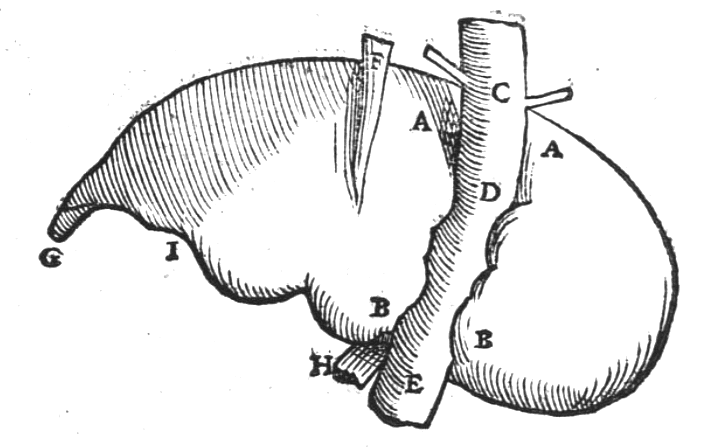
Spiegel’s lobe (lobus Spigelii) is a small posterior projection of the liver, situated between the inferior vena cava and the ligamentum venosum. First textually described by Adriaan van den Spiegel in De humani corporis fabrica (1627), although it had earlier appeared in Tabulae anatomicae (1552) by Bartolomeo Eustachi (1510–1574)
Praeterea postico loco, prope venam cavum, lobulus quidam hepatis cernitur.
Furthermore, in the posterior position, near the vena cava, a certain small lobe of the liver is seen.
Spigelii, 1627
See Figure XVIII (Decimaoctava quinti libri) identified as D, E – Hoc intervallo sedes notatur, qua cavae caudex posteriori iecoris sedi innascitur → this interval marks the site where the trunk of the vena cava is joined to the posterior surface of the liver
Spiegel’s lobe was later termed the caudate lobe (lobus caudatus) by anatomists, and reclassified as ‘segment 1’ by the French surgeon-anatomist, Claude Couinaud (1922–2008) in his liver segmentation system in 1954
The lobe was termed “lobus Spigelii” in the anatomical texts such as Johann Vesling, Syntagma Anatomicum, 1641, and Govard Bidloo, 1685 and by the 18th century, it was consistently referred to as Spiegel’s lobe in anatomical teaching.
Spigelia (Plant Genus)
Named in Spiegel’s honour. Around 60 species in Loganiaceae. Spigelia marilandica roots/rhizomes were traditionally used as an anthelmintic against intestinal parasites.
Controversies
Alternate names: Adrianus Spigelius; Adriani Spigelii; Adriano Spigeli; Spigel; Spieghel; Spiegelius
Major Publications
- Adriani Spigelii Bruxellensis philosophi ac med. pat. Isagoges in rem herbariam libri duo. Ad illustrissimam quae patavii est germanicam nationem 1606
- Spiegel A. De humani corporis fabrica libri decem. 1627 (Posthumously)
- Liber III, Caput IV–VI, [Spigelian line]
- Liber V, Fig. XVIII [Spiegel’s Lobe]
- Spiegel A. Isagoges in rem herbariam libri duo Adrianus Spigelius. 1633 (Posthumously)
- Spiegel A. Adriani Spigelii Bruxellensis. 1645 (Posthumously)
- Spiegel A. Adriani Spigelii Brvxellensis Eqvitis. 1645 (Posthumously)
- Spiegel A. The workes of that famous chirurgion. 1649 (Posthumously by Thomas Johnson)
References
Biography
- Marinus, JR. Éloge de A. van den Spieghel: lu dans la séance publique annuelle de l’Académie Royale de Médecine de Belgique, le 29 novembre 1846
- Lindeboom GA. Adriaan van den Spiegel (1578-1625). 1978
- Ghosh SK, Sharma S, Biswas S, Chakraborty S. Adriaan van den Spiegel (1578-1625): anatomist, physician, and botanist. Clin Anat. 2014 Oct;27(7):952-7.
- Dunea G. Adrianus Spigelius, the last great Paduan anatomist. Hektoen International 2019
- Bibliography. Spiegel, Adriaan van den 1578-1625. WorldCat Identities
Eponymous terms
- Klinkosch JT. Programma quo divisionem herniarum, novamque herniae ventralis speciem proponit, nec non anatomicas sectiones, et demonstrationes publicas hyemales anni MDCCLXIV… Pragae: litteris Joannis Josephi Clauser; 1764. 36 p
- Joseph Thaddaeus Klinkosch Programma, quo divisionem herniarum, novanaejue hemiae ventral is speciem proponit. Pragae 1764
- Couinaud C. Lobes et segments hépatiques: notes sur l’architecture anatomiques et chirurgicale du foie [Liver lobes and segments: notes on the anatomical architecture and surgery of the liver ]. Presse Med (1893). 1954 May 5;62(33):709-12.
- Couinaud C. Le foie; études anatomiques et chirurgicales. Masson, 1957
- Foucou B, Ould Said H, Joyeux A, M’Jahed A, Saint-Aubert B, Hamdouch Z, Joyeux H, Solassol C. Le segment I du foie ou lobe de Spigel. Etude anatomique et intérêt chirurgical [Segment I of the liver or spigelian lobe. Anatomic study and surgical value]. J Chir (Paris). 1983 Mar;120(3):179-86.
- Spangen L. Spigelian hernia. World J Surg. 1989 Sep-Oct;13(5):573-80.
- Skandalakis PN, Zoras O, Skandalakis JE, Mirilas P. Spigelian hernia: surgical anatomy, embryology, and technique of repair. Am Surg. 2006 Jan;72(1):42-8.
- Polistina FA, Garbo G, Trevisan P, Frego M. Twelve years of experience treating Spigelian hernia. Surgery. 2015 Mar;157(3):547-50.
- Ussia A, Imperato F, Schindler L, Wattiez A, Koninckx PR. Spigelian hernia in gynaecology. Gynecol Surg. 2017;14(1):8
- Laios K, Mavrommatis E, Kostoulas G, Manes K, Lagiou E, Lytsikas-Sarlis P, Piagkou M. Adrianus Spigelius’ (1578 – 1625) Ocular Anatomy. Acta Med Acad. 2019 Aug;48(2):250-254.
- Buttner R, Lee J. De-eponymising anatomical terminology. 2020
- van Gulik TM. The caudate lobe described by Adriaan van den Spiegel. Hepatobiliary Surg Nutr. 2021 Dec;10(6):748.
- Shtanko Y, Galtes J, Mata W. Spigelian Hernia: A Rare Ventral Hernia. Cureus. 2024 Feb 29;16(2):e55209.
Eponym
the person behind the name
BA MA (Oxon) MBChB (Edin) FACEM FFSEM. Emergency physician, Sir Charles Gairdner Hospital. Passion for rugby; medical history; medical education; and asynchronous learning #FOAMed evangelist. Co-founder and CTO of Life in the Fast lane | On Call: Principles and Protocol 4e| Eponyms | Books |