
![]()
Ballooning Up
aka Pulmonary Puzzler 006
A 36 year-old immunosuppressed male was infected with swine-origin influenza virus (SOIV, the 2009 H1N1 pandemic influenza A virus). He had required 3 weeks of mechanical ventilation for ‘FLAAARDS’ (‘flu’ A-associated acute respiratory distress syndrome) resulting in type 1 and 2 respiratory failure, which was complicated by bilateral pneumothoraces. These were treated with bilateral intercostal catheters (ICCs). Overnight the inspiratory pressures needed to maintain his tidal volume had progressively increased and his face had become markedly swollen.
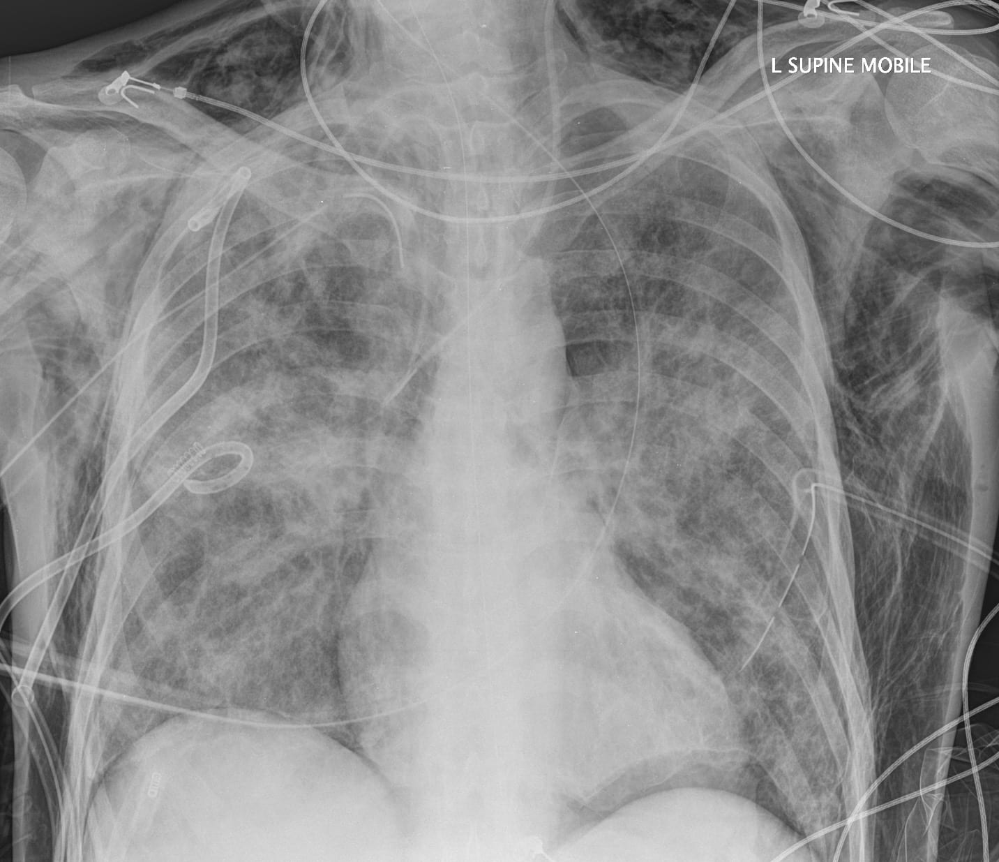
A chest radiograph was performed:
Questions
Q1. Describe the chest radiograph
Answer and interpretation
Chest radiograph findings:
- supine film.
- tracheostomy tube, right internal jugular central venous catheter, and a nasogastric tube are all visible and appropriately positioned.
- bilateral infiltrates consistent with ARDS.
- bilateral ICCs are present (somewhat kinked on the left), as well as two pigtail ‘pneumocath’ tubes on the right side.
- although there is no pneumothoraces visible on this supine film there is widespread severe subcutaneous emphysema. The subcutaneous emphysema extends up the neck and along both upper limbs. There is also a rim of air below the diaphragm, particularly between the left hemidiaphragm and the stomach.
Subcutaneous emphysema is usually the result of a pneumothorax, but can also result from surgical procedures (such as gas insufflation during laparoscopy), esophageal rupture or a pneumomediastinum.
Q2. What findings would you expect on clinical examination?
Answer and interpretation
Findings may include:
- Diffuse swellingof the involved regions:
- usually the chest and neck, but extending to the head, upper limbs and abdomen in this case.
- On palpation there may be a crackling sensation (may be absent if the subcutaneous gas is trapped locally).
- Breath sounds will be diminished.
- There may also be evidence of ‘tensioning‘:
- cyanosis due to hypoxemia, decreased chest wall movement with ventilation, neck vein distention, tachycardia and hypotension. Unchecked, this results in death.
Q3. What immediate management is indicated?
Answer and interpretation
Pneumothoraces can usually be treated with 14-20 French gauge ICCs, often using a Seldinger technique. However, in this case, bilateral large bore ICCs were inserted as the air leak is already exceeded the capacity of multiple tubes.
As marked pneumothoraces are not visible, air may be leaking around the tubes at the sites of ICC insertion. An attempt at sealing these sites with sutures and occlusive dressings is appropriate. However, it is not possible to close the pleural lining so these measures may not prevent the ongoing egress of air subcutaneously.
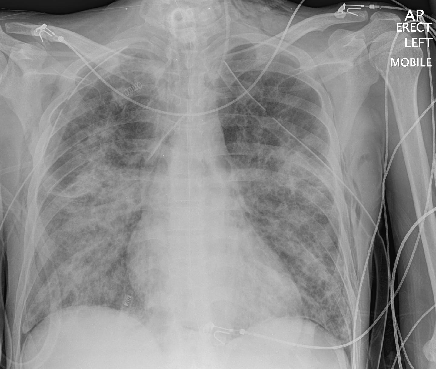
A repeat chest radiograph taken two days after the above treatment measures is shown:
Q4. What measures may help prevent a recurrence?
Answer and interpretation
Avoid barotrauma.
- Use a mechanical ventilation strategy that maintains adequate oxygenation (e.g. PaO2 >55 mmHg) and avoids excessive acidemia (e.g. keep pH >7.1) but allows permissive hypercapnia (with a tidal volume of 6-8 mL/kg) and minimises barotrauma (aim for plateau pressures <30 cmH2O).
- In refractory cases high frequency oscillatory ventilation (HFOV) or extracorporeal membrane oxygenation (ECMO) may be considered.
Ensure adequate insertion and functioning of ICCs.
- avoid making an excessive hole on ICC insertion (should be large enough to admit a finger).
- ensure all ICC side port openings are within the pleural space.
- rotate the ICC through 360 degrees after insertion to decrease the risk of tube kinking.
- try to obtain an adequate seal around the ICC insertion sites: close the wounds with sutures, seal with petroleum gauze and apply an occlusive dressing. In the patient ventilated with postive pressure this is not critical as air cannot leak in – in fact a leak around the ICC may help prevent tensioning should the ICC stop draining adequately.
- persistent air leaks may require the insertion of multiple ICCs (e.g. bronchopleural fistulae).
- apply suction initially (e.g. -20 cmH20 at the wall) – but remember that the amount of suction in the ICC is dependent on the depth of water in the water seal reservoir, not on the suction from the wall valve.
- monitor chest drains to ensure they are still bubbling or ‘swinging’.
Monitor closely for evidence of pneumothorax and subcutaneous emphysema.
Refractory air leaks may require surgical intervention:
- this is usually considered after 72 hours of persisting air leak or if lung re-expansion has not occurred.
- Options range from bronchoscopic repair using sealing agents (e.g., fibrin glue, albumin-glutaraldehyde tissue adhesive, stents, metallic coils, bone, absolute alcohol, Nd:YAG laser) to thoracostomy and lobectomy.
References
- For more Pulmonary cases check out the LITFL Top 150 Chest X-Rays
CLINICAL CASES
Pulmonary Puzzler
Intensivist in Wellington, New Zealand. Started out in ED, but now feels physically ill whenever he steps foot on the front line. Clinical researcher, kite-surfer | @DogICUma |

Need to remove HFOV from the answers for at least 2 case studies I’ve seen so far, including this one. In this case HFOV may increase the risk of worsening PTX/air leak. At any rate there isn’t any evidence for the use of HFOV in the adult population. VV ECMO should probably be added in in as an option for severe cases.