Bladder Stone: Case 1
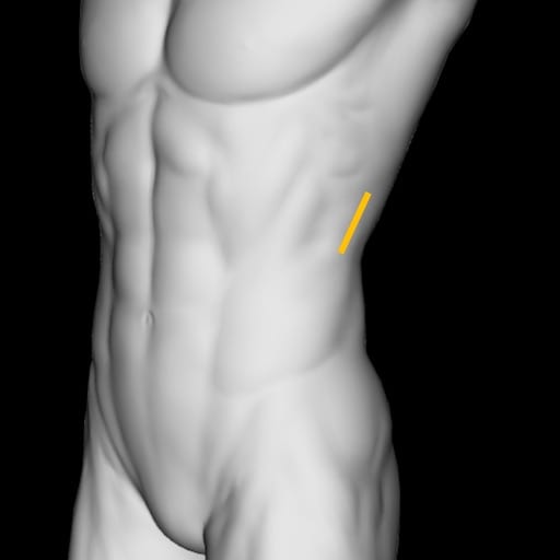
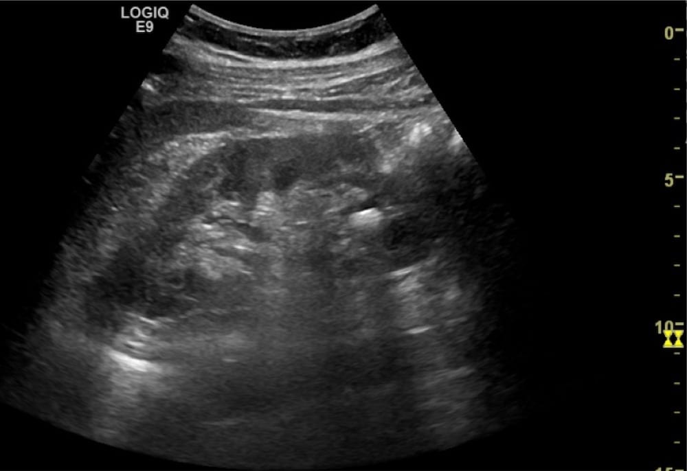
This patient is presenting with 1 day of left flank pain that resolved in the emergency department. What does this image show? Does it explain the patient’s pain?
Reveal Answer
- This is an image of the left kidney in longitudinal section without hydronephrosis.
- In the inferior pole there is a hyperechoic structure with posterior acoustic shadowing consistent with an intrarenal stone.
- Intrarenal stones do not cause symptoms. Pain is caused by ureteric spasm and inflammation as the calculus makes its way to the bladder.
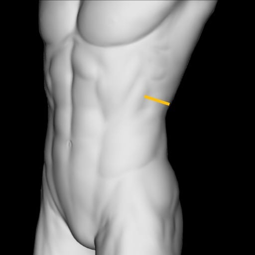
How was the probe manipulated to acquire the image below?
Reveal Answer
The probe was rotated counter clockwise using the stone as a fulcrum and now the stone can be seen in the inferior pole of the kidney cut in transverse section.
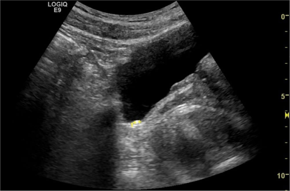
What does the image below show?
Reveal Answer
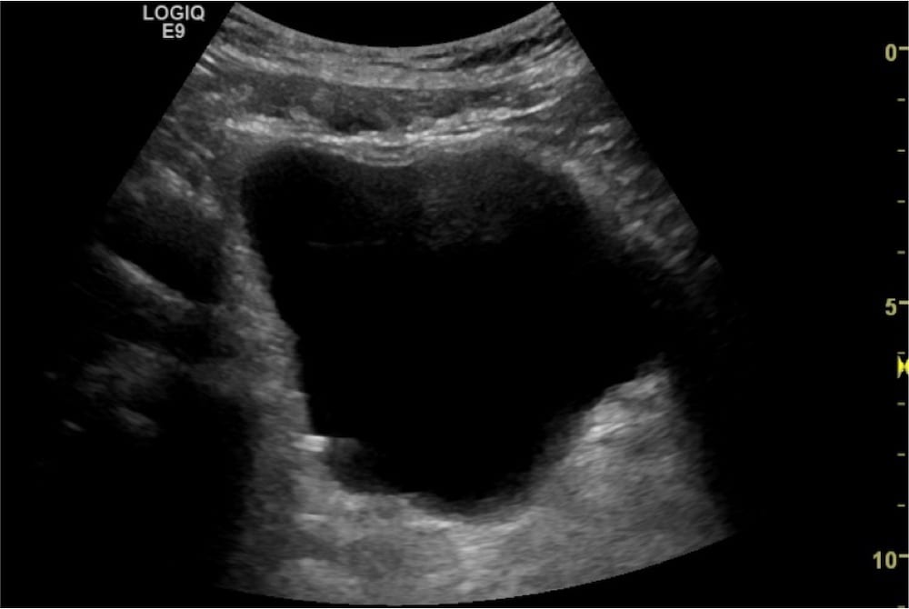
- This is a longitudinal section of the bladder with a hyperechoic, shadow casting structure resting on the posterior wall of the bladder.
- This most likely represents a calculus within the bladder.
How was the patient moved to acquire the image below and confirm this is a bladder calculus?
Reveal Answer
- The patient was placed in a right decubitus position and a transverse image of the bladder was taken.
- The bladder calculus is now on the right posterior bladder wall, confirming it is free floating.
- This stone likely just passed into the bladder as the patients symptoms resolved.
Related Clinical Cases
- LITFL Ultrasound library
- RENAL ultrasound MODULES
- LITFL Top 100 ultrasound cases
- RENAL ultrasound WORKED CASES
ULTRASOUND LIBRARY
Clinical Cases
An Emergency physician based in Perth, Western Australia. Professionally my passion lies in integrating advanced diagnostic and procedural ultrasound into clinical assessment and management of the undifferentiated patient. Sharing hard fought knowledge with innovative educational techniques to ensure knowledge translation and dissemination is my goal. Family, wild coastlines, native forests, and tinkering in the shed fills the rest of my contented time. | SonoCPD | Ultrasound library | Top 100 | @thesonocave |

