![]()
Charles Lockwood
Charles Barrett Lockwood (1856-1914) was an English surgeon.
Research interests in morphological anatomy making a significant contribution to the understanding of eye anatomy, describing Lockwood’s suspensory ligament and Lockwood’s tendon in 1885.
Lockwood is remembered for his surgical work with femoral and inguinal hernias and his infra-inguinal (Low) approach in elective femoral hernia repair (Lockwood operation, 1898)
Also made contributions in the field of bacteriology, asepsis and aseptic surgical techniques. Ironically died as a result of sepsis following intra-operative needle-stick injury in 1914
Biography
- Born September 23, 1856 Stockton-On-Tees, England
- 1881 – FRCS, Fellow of the Royal Colleges of Surgeons
- 1882 – Surgeon to the Great Northern Central Hospital
- 1886-1889 Hunterian Professor of Comparative Anatomy and Physiology
- 1887 – Founded the British Anatomical Society
- 1892 – Assistant Surgeon to St. Bartholomew’s Hospital
- 1894-1895 Hunterian Professor of the Royal College of Surgeons of England
- 1903 – Consulting Surgeon to St. Bartholomew’s Hospital
- 1912 – Resigned from active staff at St. Bart’s but continued to work as Consulting Surgeon and Governor of the hospital
- President of the Anatomical Society (1902); President of the Medical Society of London; and the Harveian Society of London.
- Died November 8, 1914 from septicaemia after sustaining a pin-prick injury when performing an appendicectomy
Medical Eponyms
Lockwood’s operation (1898)
Infra-inguinal approach (low approach) for femoral hernia repair. Lockwood’s infra-inguinal approach is the preferred method for elective repair of uncomplicated femoral hernia. The approach is through an oblique incision 1cm below and parallel to the inguinal ligament. [Radical cure of femoral hernia 1898: 176-206]
Lockwood sign (1932)
In a letter to the Editors of British Medical Journal, George Herbert Colt (1878-1957) wrote that the late Mr. CB. Lockwood taught his dressers as follows:
The patient lies on his back with his head raised on a pillow and his knees drawn up, so that the superficial abdominal muscles are relaxed. The surgeon sits down near his right side and palpates the right iliac region near McBurney’s spot with the three inner fingers of his left hand. If he feels a trickle of flatulence passing his fingers, and if this can be often repeated after waiting a half to one minute or a little longer, the patient has either a chronically inflamed appendix or adhesions near it.
Colt. 1932
Mr Colt further refined the definition of Lockwood sign and described his experience of it:
The trickle of flatulence was expressly stated not to be the large gurgle of gas collected in the caecum, but a small, fine crepitation with a higher note. During the last ten years I have tested this sign in over 600 cases verified by operation; it has, in fact, been the determining sign. It has been positive in every case but one.
Colt. 1932
Lockwood’s suspensory ligament of the eye (1885)
Lockwood described the muscle-associated connective tissue in the inferior orbit, thickening of the Tenon capsule and the suspensory ligament:
a band of fibrous tissue, stretched, like a sling, from one side of the orbit to the other…inserted into the malar and the lachrymal bones…intimately woven with the capsule of Tenon, but not to such an extent as to conceal the identity of its fibres. (Fig 4 and 5)
Lockwood 1885: 16

Fig. 4. Vertical section. The sheath of the levator palpebae is dotted in above the superior rectus. The capsule of Tenon is thicker below in the region of the suspensory ligament. (I. 0) inferior oblique. Only the tunica vaginalis is delineated; the tunica adventitia has been omitted.
Fig. 5. Horizontal section through the orbit at the level of the canthi. (L) lachrymal sac. The front aperture is crossed by the suspensory ligament, which is pierced by the inferior rectus and inferior oblique.
Lockwood’s tendon of the eye (1885)
Lockwood described the common origin of the superior rectus, the upper half of the medial, and the superior head of the lateral rectus muscles; this structure is sometimes called “Lockwood’s tendon.” Lockwood discovered that the superior rectus has:
The optic nerve and its sheath together with loose connective tissue having been removed, it became evident that the under surface of the superior rectus possessed a tendon very similar to that of the inferior rectus. The structure is fastened to the upper and outer margin of the optic foramen. Anteriorly it is aponeurotic, and gives attachment to the superior rectus, the upper part of the internal rectus, and the superior head of the external rectus. The fasciculus, which belongs to the latter muscle, passes down its posterior border, and is continuous with the slip which this muscle receives from the tendon of Zinn (Fig 2)
Lockwood 1885: 3
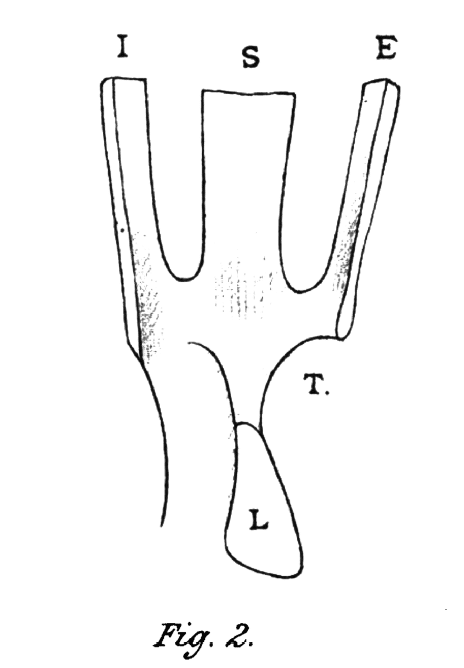
Fig 2: Common tendinous origin of superior (S), external (E), and internal (I) recti. (L) root of the lesser wing of the sphenoid, to the front of which it is attached (T) tendon.
Notable Quotables
Perhaps the day will arrive when a man of genius will invent some method for bringing the soft tissues of the body within the range of our vision, just as the X-rays have brought the bones into sight.
Lockwood 1911
Major Publications
- Bruce-Clark W, Lockwood CB. The dissector’s manual. 1883
- Lockwood CB. I. On abnormalities of the caecum and colon with reference to development. St Bartholomew’s Hospital Reports, 1884; XIX
- Lockwood CB. The development of the great omentum and transverse mesocolon. 1884
- Lockwood CB. The Anatomy of the Muscles, Ligaments, and Fasciae of the Orbit, including an Account of the Capsule of Tenon, the Check Ligaments of the Recti, and the Suspensory Ligaments of the Eye. Journal of anatomy and physiology. 1885; 20(1): 1-25. [Lockwood’s suspensory ligament] [Lockwood’s tendon]
- Lockwood CB. The development of the arteries of the abdomen and their relation to the peritoneum. Proceedings of the Royal Society. 1885; 238
- Lockwood CB. Development and transition of the testis: normal and abnormal. Hunterian Lecture. 1888
- Lockwood CB. The early development of the pericardium, diaphragm, and great veins. Philosophical Transactions of the Royal Society of London. 1888; 179: 365-384
- Lockwood CB. Morbid anatomy, pathology, and treatment of hernia. Hunterian Lecture. 1889
- Lockwood CB. On the fossae round the caecum, and the position of the vermiform appendix: with special reference to retro-peritoneal hernia. 1892
- Lockwood CB. The radical cure of hernia, hydrocele, and varicocele. 1898 [Lockwood operation 176-206]
- Lockwood CB. Appendicitis: its pathology and surgery. 1901
- Lockwood CB. Aseptic surgery in theory and practice. 1904
- Lockwood CB. Clinical surgery. 1911
References
- In memoriam Charles Barrett Lockwood, 1856-1914. Saint Bartholomew’s Hospital reports. 1914
- Charles Barrett Lockwood, F.R.C.S. Br Med J. 1914; 2(2812): 903-904
- Charles Barrett Lockwood. J Anat Physiol. 1915; 49(2): 240–242.
- Colt GH. Chronic Appendicitis: “Lockwood’s Sign”. Br Med J. 1932 Nov 19; 2(3750): 942.
- Obituary: George Herbert Colt (1878-1957). Br Med J 1957; 2: 1118
- Dahlmann-Noor AH. The Origins and Insertions of Rectus Muscles: Paul J. Tillaux and Charles B. Lockwood. Strabismus. 2008; 16(4): 170-3.
- Rastogi V et al. Abdominal Physical Signs and Medical Eponyms: Part III. Physical Examination of Palpation, 1926–1976. Clin Med Res. 2019; 17(3-4): 107-114.
- Bibliography. Lockwood, Charles Barrett 1856-1914. WorldCat Identities
- Cadogan M. Eponymous signs in Appendicitis. LITFL
Eponym
the person behind the name
BA MA (Oxon) MBChB (Edin) FACEM FFSEM. Emergency physician, Sir Charles Gairdner Hospital. Passion for rugby; medical history; medical education; and asynchronous learning #FOAMed evangelist. Co-founder and CTO of Life in the Fast lane | On Call: Principles and Protocol 4e| Eponyms | Books |