
![]()
COPD Case 2 Lung Bullae
A patient with a history of COPD / severe emphysema presents with an exacerbation of their shortness of breath.
What does this ultrasound clip demonstrate?
Reveal Answer
This clip is taken with the linear ultrasound transducer placed directly over a lung bullae.
There is complete loss of lung sliding and there are no short path reverberation artefacts (B-lines or comet tails).
The appearance is essentially identical to that of a pneumothorax and is a potential major pitfall of ultrasound in this setting.
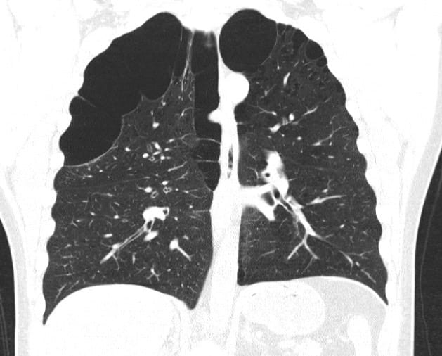
A CT scan of the chest is performed. Describe the CT and the implications in ultrasound scanning.
Reveal Answer
This patient has severe emphysema with bullae formation.
Whilst they are at risk of developing a pneumothorax, differentiating pneumothorax from bullous emphysema is often very difficult with ultrasound and alternate imaging is recommended.
A chest x-ray may clarify any doubt but CT is often required.
Related Clinical Cases
- LITFL Ultrasound library
- LITFL TOP 100 ultrasound cases
- LUNG ultrasound WORKED CASES
- LUNG ultrasound LEARNING MODULES

ULTRASOUND LIBRARY
Clinical Cases

An Emergency physician based in Perth, Western Australia. Professionally my passion lies in integrating advanced diagnostic and procedural ultrasound into clinical assessment and management of the undifferentiated patient. Sharing hard fought knowledge with innovative educational techniques to ensure knowledge translation and dissemination is my goal. Family, wild coastlines, native forests, and tinkering in the shed fills the rest of my contented time. | SonoCPD | Ultrasound library | Top 100 | @thesonocave |