![]()
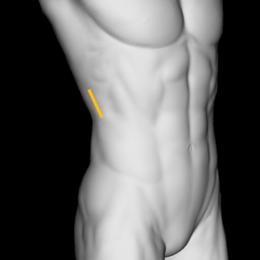
Distal Ureteric Stone: Case 1
This patient presents with 24 hours of right flank pain locaslised to the right lower abdomen.
What does this image of the right kidney in longitudinal section show?

Reveal Answer
- The superior pole of the kidney (screen left) is obscured by rib posterior acoustic shadow artefact. An additional image is required to show the superior pole of the kidney.
- There is a prominent renal pelvis and mild hydronephrosis present.
What does this image of the right kidney in transverse section show?
Reveal Answer
- The probe has lost contact with the skin at the posterior side of the kidney (screen left), and attempts should be made to correct this loss of contact.
- This image shows the mild hydronephrosis and prominent pelvis in transverse section.
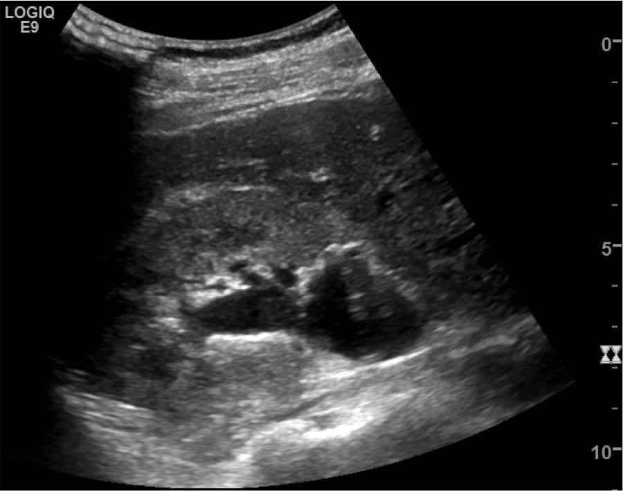
What does the image below show, and how was it obtained?
Reveal Answer
- The image is of the right proximal hydroureter (>3mm) connecting with the renal pelvis in long.
- This image is obtained by acquiring the previous image of the right kidney in longitudinal section. Then, either fanning or moving the probe posteriorly and caudally while slightly rotating the probe counter clockwise. It is important to keep the pelviureteric junction in the centre of the screen to eventually open up the longitudinal image of the ureter and renal pelvis seen above.
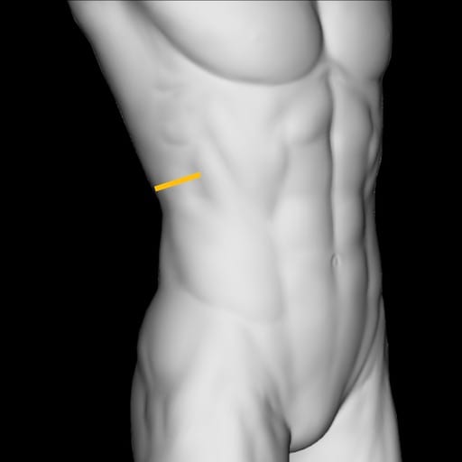
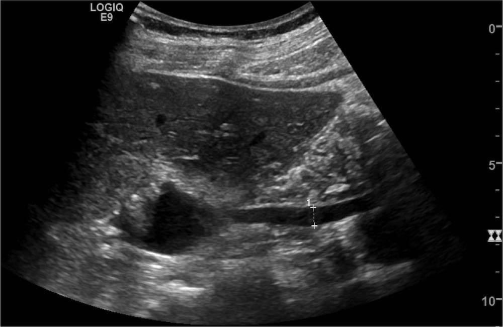
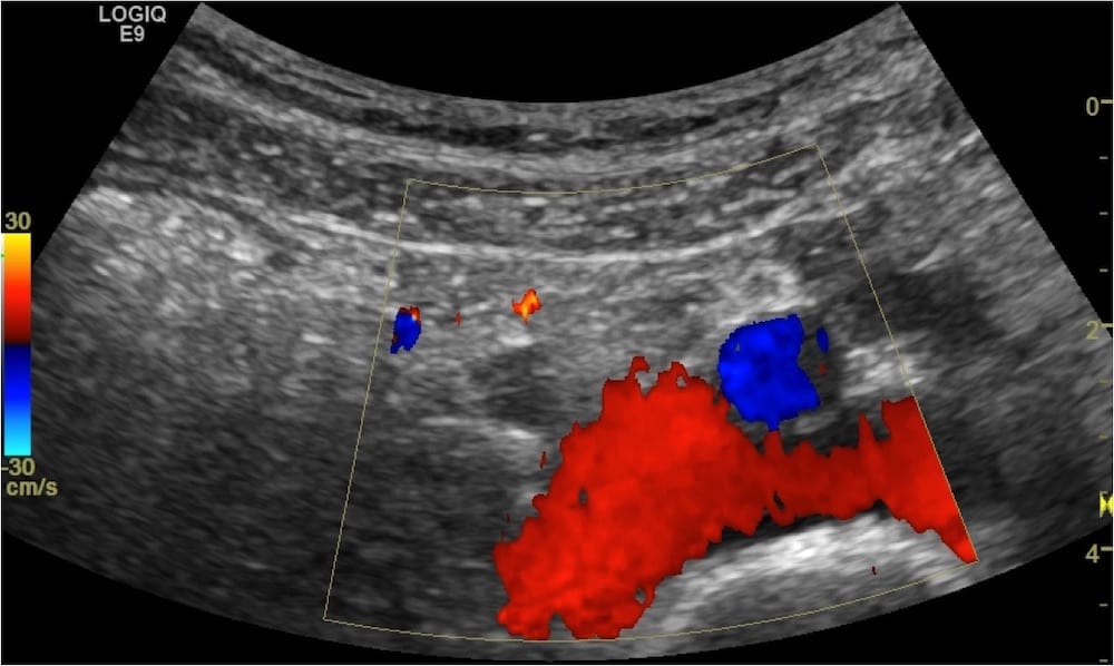
The hydroureter can be commonly seen at the pelvic brim, what does this transverse image at the right pelvic brim show?
Reveal Answer
- This is a colour Doppler image showing the iliac vessels in blue and red, with a transverse image of the hydroureter, the circular structure not highlighted by colour, going over the top of the vessels. A common place for calculi to obstruct.
- A normal ureter without distension is usually not able to be visualised.
- This image informs us that the area of obstruction is distal to this point.
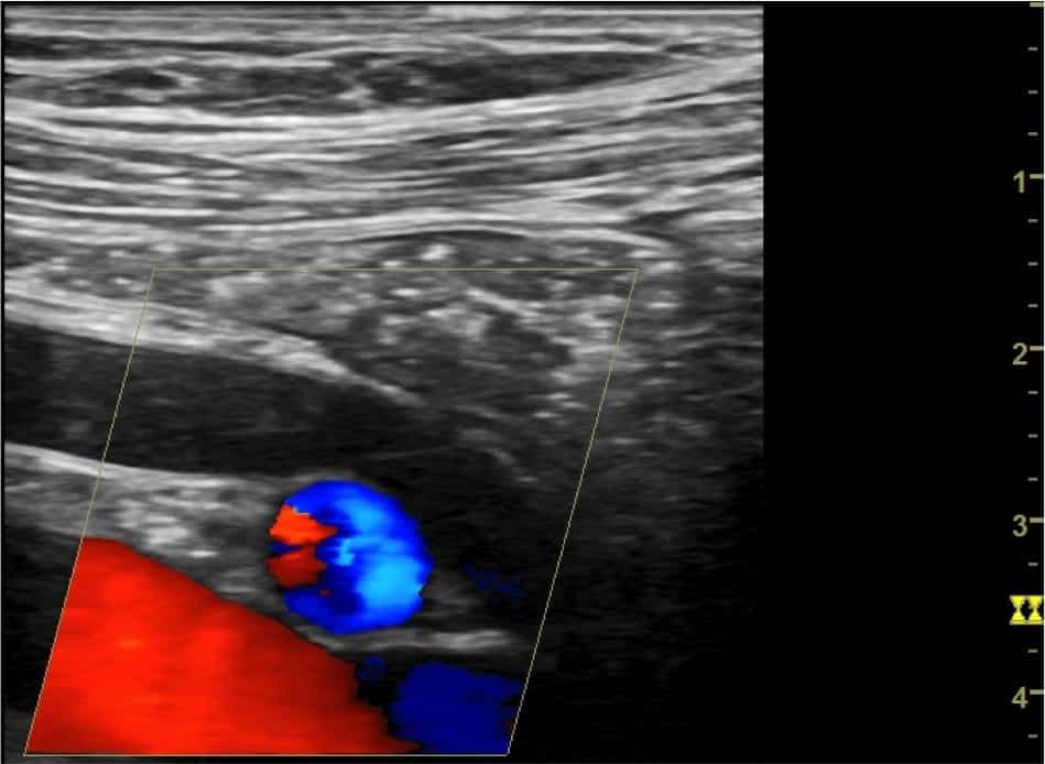
How was the previous image manipulated to acquire this image?

Reveal Answer
- The probe was changed to a linear probe to increase frequency and enhance image resolution.
- After visualising the iliac vessels and finding he ureter in transverse, the probe is rotated clockwise with the ureter in the centre to open up a longitudinal image.
- This image is useful to follow the ureter as it dives into the pelvis in order to find the obstructing calculus for measurement.
- The calculus was not visualised here or at the distal ureter/vesicoureteric junction. As long as there is no complicating factors (i.e. acute kidney injury, single kidney, urine infection, prolonged symptoms, able to tolerate oral fluids, concern for alternate diagnosis, etc), this may be managed conservatively as an outpatient and reconsider repeat imaging if symptoms do not resolve.
Related Clinical Cases
- LITFL Ultrasound library
- RENAL ultrasound MODULES
- LITFL Top 100 ultrasound cases
- RENAL ultrasound WORKED CASES

ULTRASOUND LIBRARY
POCUS

An Emergency physician based in Perth, Western Australia. Professionally my passion lies in integrating advanced diagnostic and procedural ultrasound into clinical assessment and management of the undifferentiated patient. Sharing hard fought knowledge with innovative educational techniques to ensure knowledge translation and dissemination is my goal. Family, wild coastlines, native forests, and tinkering in the shed fills the rest of my contented time. | SonoCPD | Ultrasound library | Top 100 | @thesonocave |