![]()
Distal VUJ stone: Case 1
This patient presented with right-sided groin pain, nausea, vomiting and gross haematuria.
What does this image show?

Reveal Answer
- This image shows the right kidney in longitudinal section, the lower pole is partially obscured by bowel gas.
- The is mild prominence of the renal pelvis, but this is within normal limits.
- There is no hydronephrosis or hydroureter.
- There are no intrarenal calculi evident.
- There is no perinephric renal fluid – a common finding in acute renal colic.
Convinced the patient has a ureteric calculus, you search the ureter for a stone.
What does this clip show?

Reveal Answer
- This clip shows the distal right ureter in longitudinal section.
- There is mild hydroureter, which can be followed down to an obstructing calculus.
- Colour Doppler is applied to demonstrate twinkle artefact of the calculus.
What does this transverse image of the bladder demonstrate?
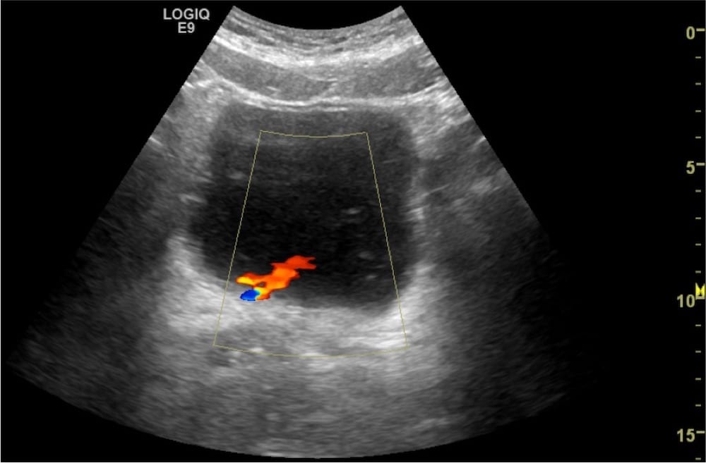
Reveal Answer
- This image shows a normal appearing right ureteric jet
- The presence of this jet with the absence of hydronephrosis means the VUJ stone is non-obstructive.
Related Clinical Cases
- LITFL Ultrasound library
- RENAL ultrasound MODULES
- LITFL Top 100 ultrasound cases
- RENAL ultrasound WORKED CASES

ULTRASOUND LIBRARY
Clinical Cases

An Emergency physician based in Perth, Western Australia. Professionally my passion lies in integrating advanced diagnostic and procedural ultrasound into clinical assessment and management of the undifferentiated patient. Sharing hard fought knowledge with innovative educational techniques to ensure knowledge translation and dissemination is my goal. Family, wild coastlines, native forests, and tinkering in the shed fills the rest of my contented time. | SonoCPD | Ultrasound library | Top 100 | @thesonocave |