![]()
Distal VUJ stone: Case 2
This patient presented with left-sided abdominal pain radiating to his testicle.
What does this image show?
Reveal Answer
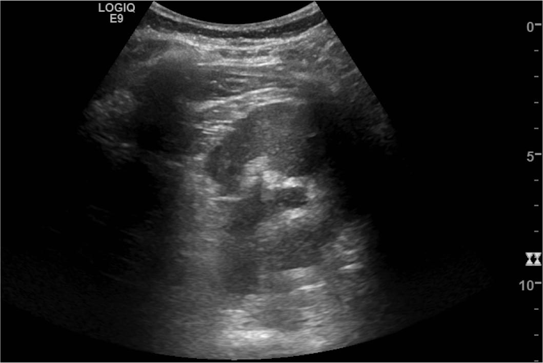
- This image shows the left kidney in longitudinal section.
- There is mild hydronephrosis.
- There is no hydroureter demonstrated in this image, although was seen on dynamic scanning.
- There are no obvious intrarenal calculi evident. The echogenic focus in the collecting system of the upper pole did not have posterior acoustic shadowing or twinkle artefact.
- There is no perinephric renal fluid – which is equivalent to the stranding seen on CT.
What does this image show?
Reveal Answer
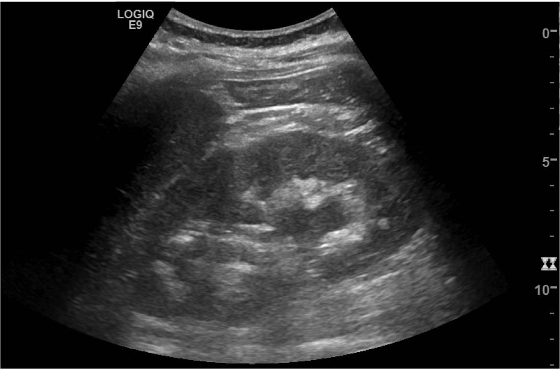
- This image shows the left kidney in a transverse section.
- There is mild hydronephrosis.
- The lateral renal cortex is obscured by rib shadow artefact.
What does this transverse image taken at the pelvic brim demonstrate?
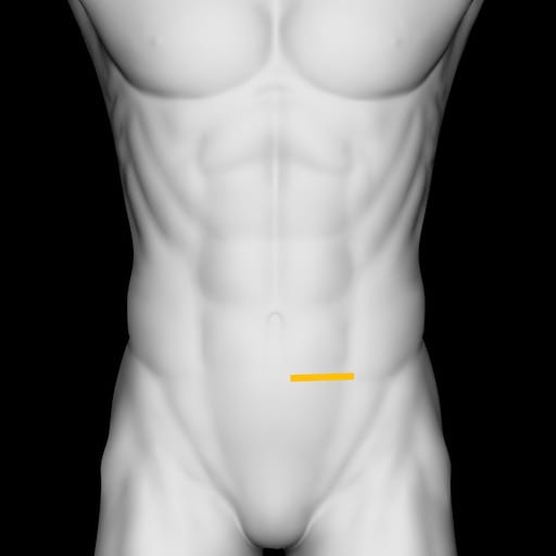
Reveal Answer
- This is a transverse section of the mid ureter at the level of the pelvic brim, where the ureter crosses the common iliac vessels.
- This is a frequent site for a ureteric calculus to obstruct. The pelvi-ureteric junction (PUJ) and ureterovesical junction (UVJ or VUJ) are the other frequent sites stones lodge.
- The normal ureter diameter is less than 3 mm, and is not normally identified at the pelvic brim. In this case mild hydroureter is demonstrated.
- Colour Doppler is used to differentiate it from a blood vessel.
What does this image demonstrate?
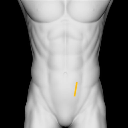
Reveal Answer
- This is a longitudinal image of the left ureter in its mid-section.
- Colour flow is again used in this image to demonstrate lack of rapid flow within the ureter.
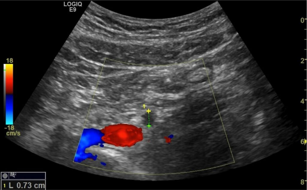
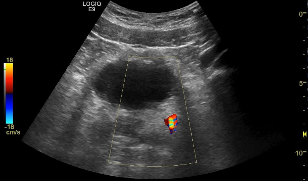
What do these transverse images of the bladder show?
Reveal Answer
- The bladder is poorly filled but appears normal.
- Posterior to the bladder, within the distal left ureter, is an echogenic calculus.
- Colour Doppler demonstrates twinkle artefact from the stone.
- Occasionally phleboliths may cause confusion. To confirm a ureteric calculus ensure the echogenic, shadow casting, calcified stone lies within the ureter, and the findings correlate clinically.
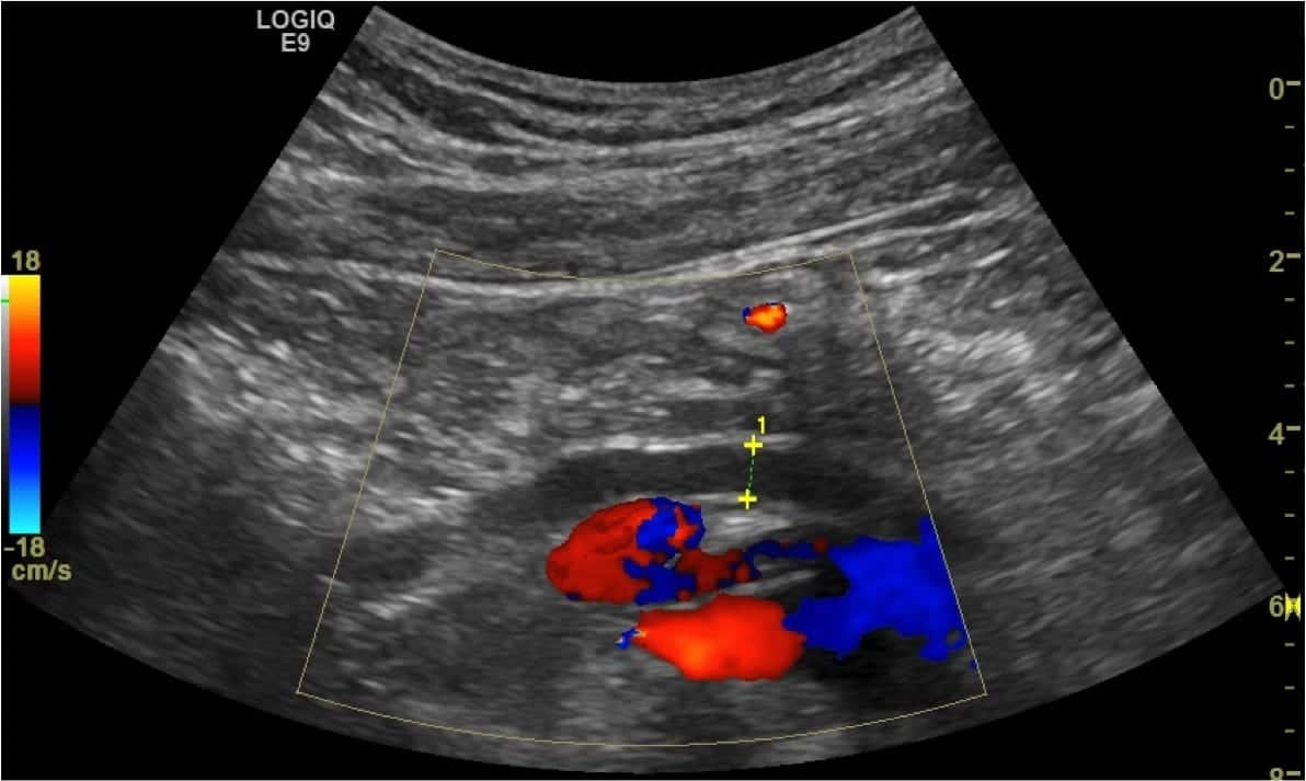
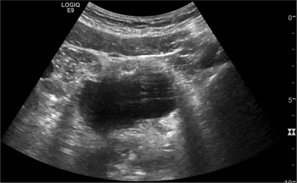
What does this longitudinal section of the bladder show?
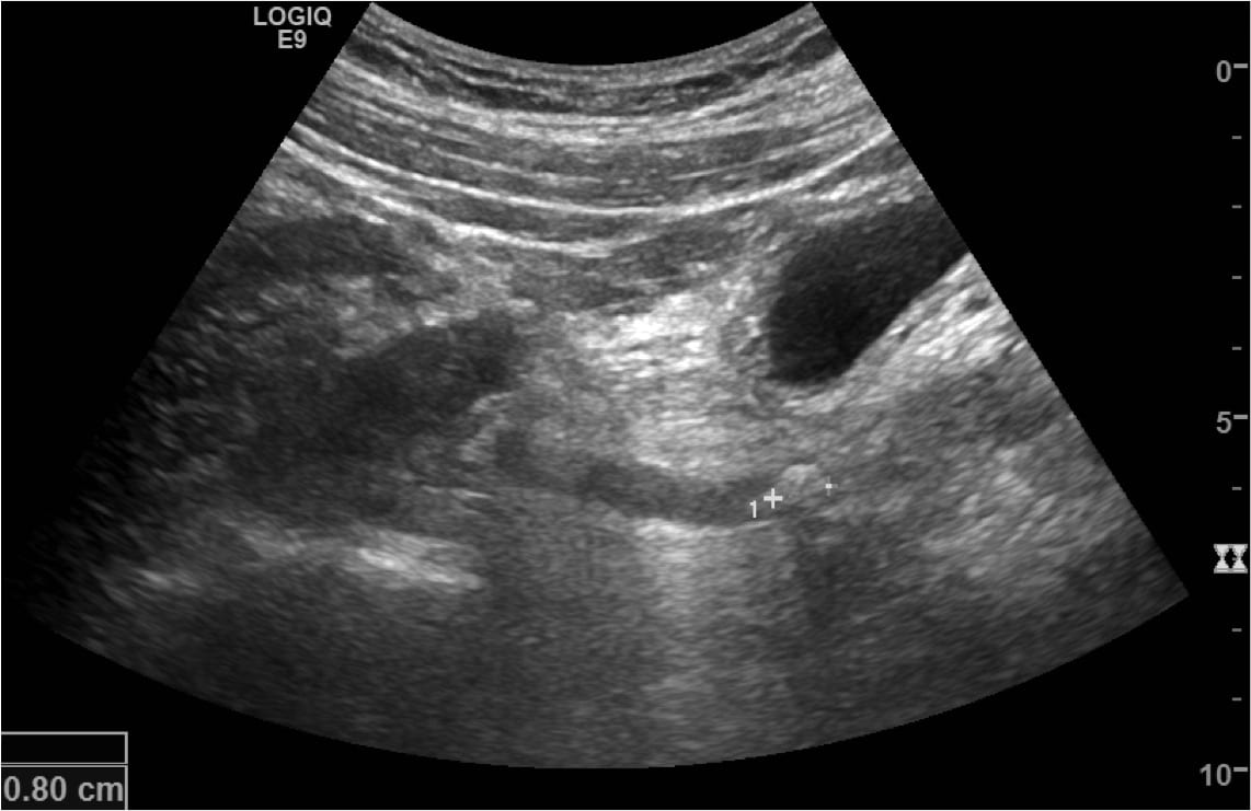
Reveal Answer
- This image shows the distended left distal ureter in longitudinal section, with the obstructing calculus toward the VUJ.
- The calculus measures 8mm, although ultrasound may slightly overestimate calculus dimensions – a function of beam-width artefact.
Related Clinical Cases
- LITFL Ultrasound library
- RENAL ultrasound MODULES
- LITFL Top 100 ultrasound cases
- RENAL ultrasound WORKED CASES

ULTRASOUND LIBRARY
Clinical Cases

An Emergency physician based in Perth, Western Australia. Professionally my passion lies in integrating advanced diagnostic and procedural ultrasound into clinical assessment and management of the undifferentiated patient. Sharing hard fought knowledge with innovative educational techniques to ensure knowledge translation and dissemination is my goal. Family, wild coastlines, native forests, and tinkering in the shed fills the rest of my contented time. | SonoCPD | Ultrasound library | Top 100 | @thesonocave |