Distal VUJ stone: Case 3
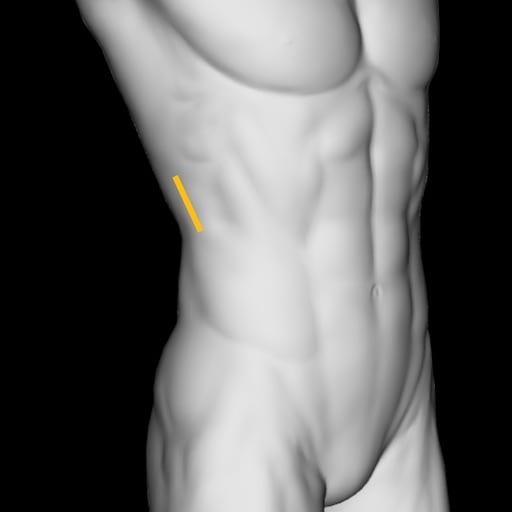
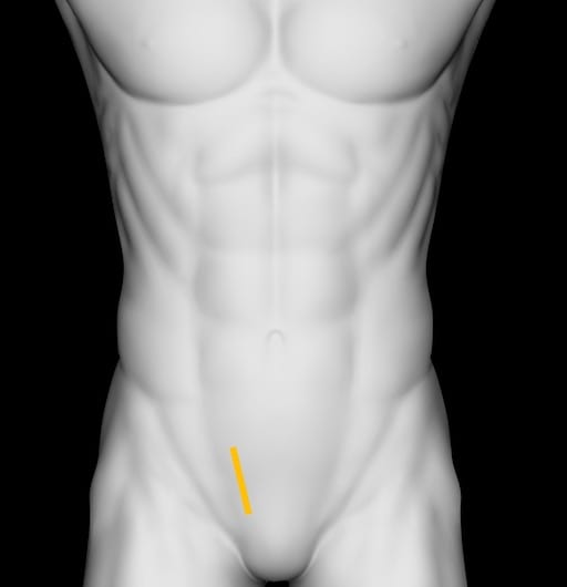
Sixty four year old female with right loin to groin pain. Urine analysis without hematuria.
What does this image show?
Reveal Answer
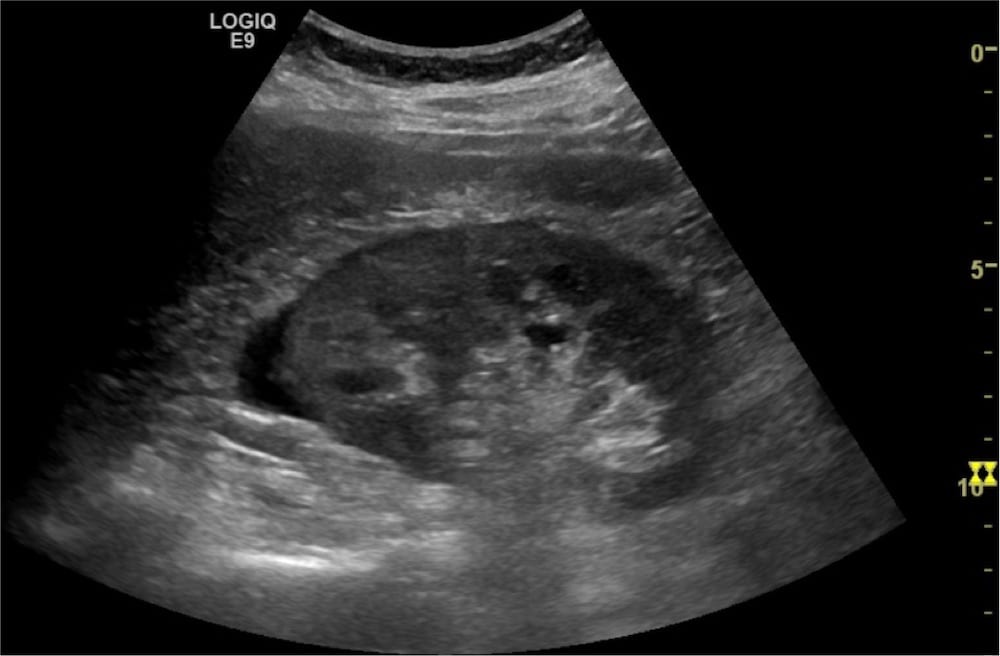
- This image shows the right kidney in longitudinal section.
- There is perinephric renal fluid at the superior pole tracking inferiorly.
- There are no intrarenal calculi.
What does this image show?
Reveal Answer
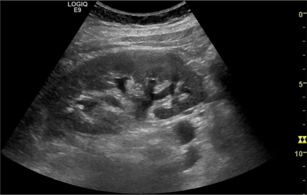
- This image shows the right kidney in a longitudinal section with the perinephric fluid tracking to the inferior pole.
- There is mild hydronephrosis.
What does this image show?
Reveal Answer
- This is a longitudinal image of the kidney with well shaped cups on the calyxes, consistent with mild hydronephrosis.
What does this transverse image of the bladder demonstrate?
Reveal Answer
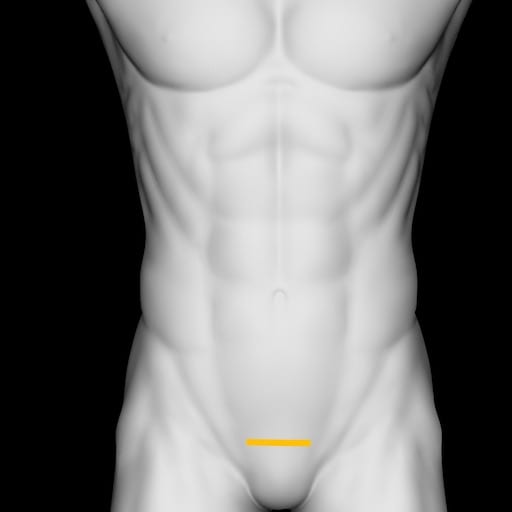
- There is an echogenic focus with posterior shadowing at the right VUJ lacking twinkle artefact. Consistent with VUJ calculi.
- This highlights that not all calculi are going to express twinkle artefact.
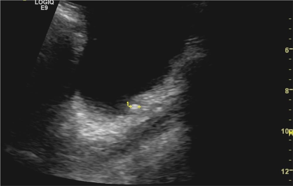
What does this longitudinal section of the bladder show?
Reveal Answer
- This image shows the bladder in a longitudinal cross section at the right VUJ.
- A calculus sit at the VUJ with posterior acoustic shadowing.
Related Clinical Cases
- LITFL Ultrasound library
- RENAL ultrasound MODULES
- LITFL Top 100 ultrasound cases
- RENAL ultrasound WORKED CASES
ULTRASOUND LIBRARY
Clinical Cases
An Emergency physician based in Perth, Western Australia. Professionally my passion lies in integrating advanced diagnostic and procedural ultrasound into clinical assessment and management of the undifferentiated patient. Sharing hard fought knowledge with innovative educational techniques to ensure knowledge translation and dissemination is my goal. Family, wild coastlines, native forests, and tinkering in the shed fills the rest of my contented time. | SonoCPD | Ultrasound library | Top 100 | @thesonocave |

