![]()
Distal VUJ stone: Case 4
Abrupt onset of right iliac fossa pain, persisting colicky pain overnight. What does this image show?
Reveal Answer
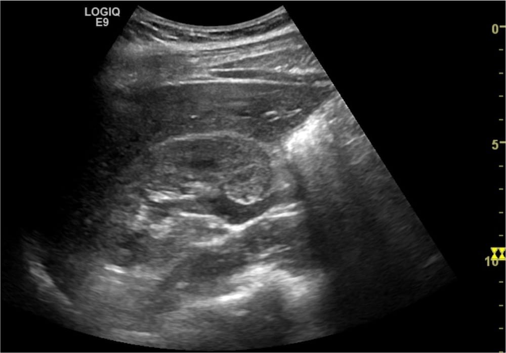
- This image shows the right kidney in longitudinal section.
- There is mild hydronephrosis and proximal hydroureter that becomes obstructed by dirty shadow from bowel gas on the right of the image.
- A thin line of perinephric fluid sits at the inferior pole.
What does this image show?
Reveal Answer
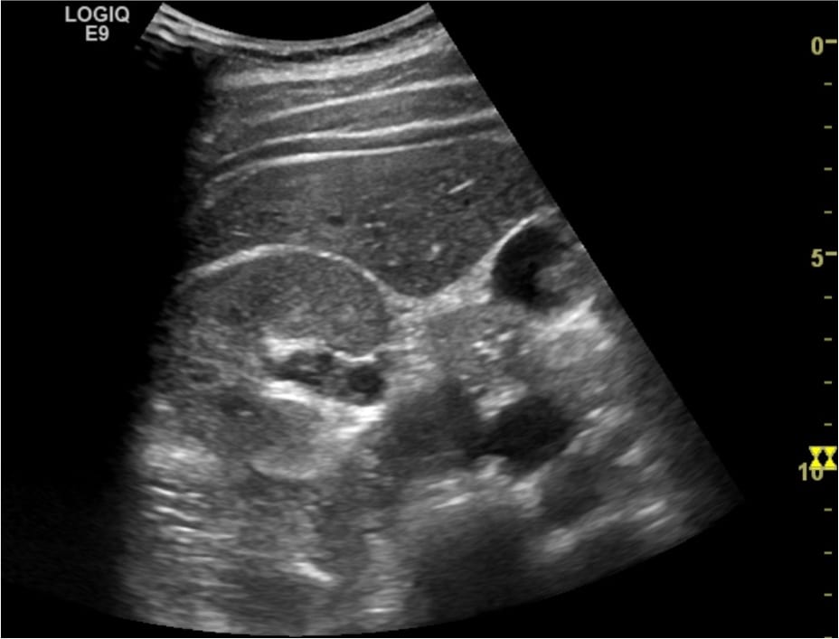
- This image shows the right kidney in a transverse section.
- There is mild hydronephrosis.
- The left side of the image drops out secondary to poor probe contact.
What does this image show?
Reveal Answer
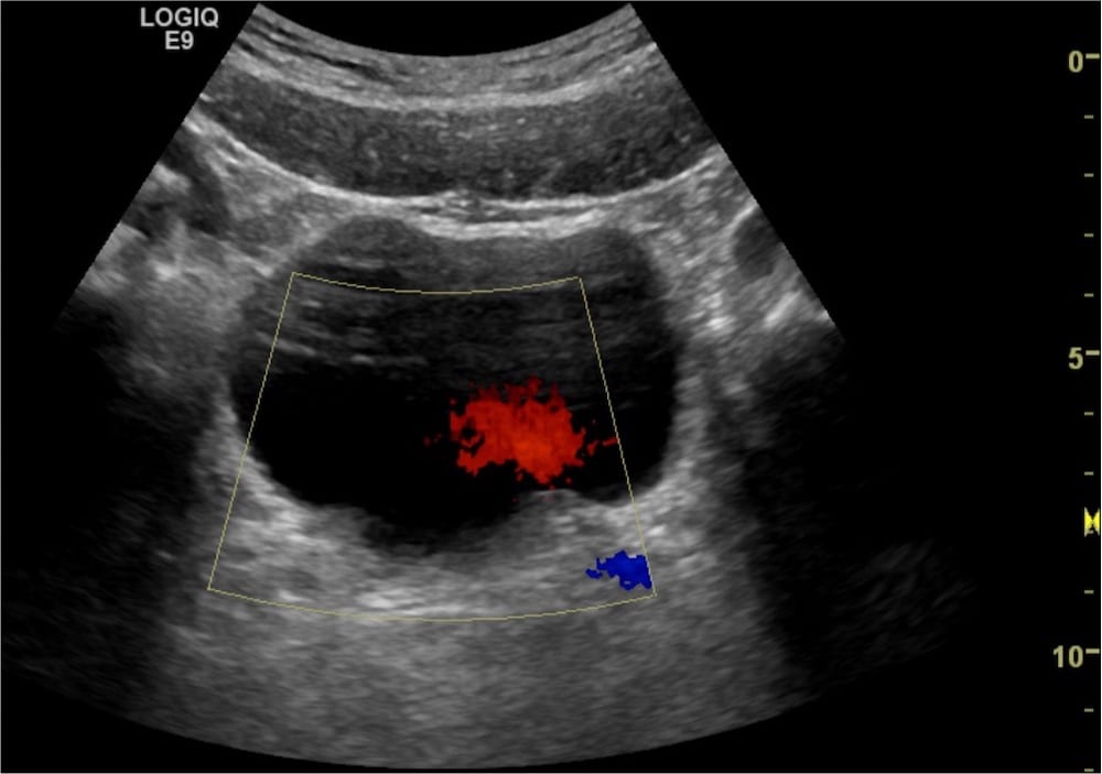
- This is a transverse image of the bladder.
- Colour Doppler at the VUJ shows a left ureteric jet.
- After search for a right ureteric jet for 2 minutes, none is demonstrated. Indicating possible right ureteral obstruction.
- Normal ureteral peristalsis occurs on average every 20 seconds.
What does this longitudinal image of the bladder demonstrate?


Reveal Answer
- There is an echogenic focus with posterior shadowing 1 cm from the right VUJ associated with hydroureter.
- TGC (Time Gain Compensation) is appropriately adjusted in this image, required for all images with posterior acoustic enhancement from cystic structures. [LINK to TGC EXPLAINATION]
What does this longitudinal section of the bladder show?

Reveal Answer
- This image shows the bladder in a longitudinal cross section at the right VUJ.
- Do not forget to utilize Twinkle Artefact when searching for a possible calculi.
Related Clinical Cases
- LITFL Ultrasound library
- RENAL ultrasound MODULES
- LITFL Top 100 ultrasound cases
- RENAL ultrasound WORKED CASES

ULTRASOUND LIBRARY
Clinical Cases

An Emergency physician based in Perth, Western Australia. Professionally my passion lies in integrating advanced diagnostic and procedural ultrasound into clinical assessment and management of the undifferentiated patient. Sharing hard fought knowledge with innovative educational techniques to ensure knowledge translation and dissemination is my goal. Family, wild coastlines, native forests, and tinkering in the shed fills the rest of my contented time. | SonoCPD | Ultrasound library | Top 100 | @thesonocave |