Funtabulously Frivolous Friday Five 172
Just when you thought your brain could unwind on a Friday, you realise that it would rather be challenged with some good old fashioned medical trivia FFFF, introducing the Funtabulously Frivolous Friday Five 172
Question 1
Which animal is winking at you?
Reveal the funtabulous answer
An owl
Spinal metastases are common in malignant tumors. Up to 80% of cancer patients have spinal metastasis at autopsy and the spine is the principle site for bony metastasis (50%).
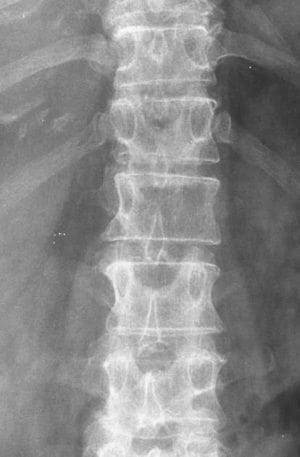
Plain XR of the spine are a valuable and readily available initial imaging study. Plain x-ray film findings include pedicle erosion, paraspinal soft-tissue shadows, wedge compression, and pathological fracture–dislocation. One of the first signs of vertebral metastases is disappearance of the pedicle on the AP X-ray. This is known as the winking owl sign.
Anteroposterior and lateral radiographs demonstrate abnormal findings in up to 90% of patients with symptomatic spinal metastasis. Lytic lesions and vertebral collapse are common; however, both osteoblastic and sclerotic alterations also occur, especially with breast and prostatic metastasis. Intervertebral disc margins are invariably spared in metastatic tumor invasion, contrasting with the disc erosion commonly observed with infectious entities.
Jacobs, 2001
The winking owl sign is a reliable sign of osteolytic spinal metastases and is classically seen on AP radiographs of the thoracolumbar spine. The loss of the normal pedicle contour and unilateral pedicle absence has been likened to that of a winking owl with the missing pedicle being the closed eye, the contralateral pedicle being the open eye and the spinous process being the beak.
In the image, the right pedicle of T8 (the left eye of T8) is not visible, suggesting destruction of the cortical bone of this pedicle. This is classically associated with an osteolytic process of metastatic disease.
Differentials include, congenital absence, neurofibromatosis, radiation therapy, spinal metastases, intraspinal malignancies, TB or lymphoma
- Jacobs WB, Perrin RG. Evaluation and treatment of spinal metastases: an overview. Neurosurg Focus. 2001;11(6)
- Cadogan M. Winking owl sign. LITFL
Question 2
Which two animals are referred to here in a patient with achalasia?
Reveal the funtabulous answer
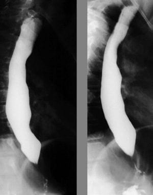
Rat’s tail and Bird’s Beak
This a sign representing the tapering of the inferior oesophagus during a barium swallow.
Question 3
Which snake can be seen here?
Reveal the funtabulous answer
The cobra head sign (or spring onion sign)
It refers to the dilatation of the distal ureter surrounded by a thin lucent line. It indicates an uncomplicated ureterocele.
If there is any irregularity or loss of definition this should raise concerns for a pseudoureterocele.
- Cobra head sign – intravenous urogram. Radiopedia
- Ultrasound Case 073. LITFL
- Tirman PA, Dyer RB. The cobra head sign. Abdom Imaging. 2015;40 (3): 609-10.
Question 4
Which four legged animal is potentially referred to in this chest X-ray?
Reveal the funtabulous answer
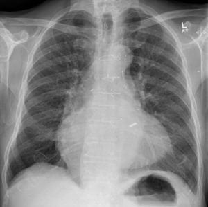
Stag’s antler sign
Upper lobe pulmonary venous diversion (cephalisation). Prominence of the pulmonary veins are said to resemble stag’s antlers, the earliest sign of pulmonary oedema (pulmonary venous hypertension)
- Stag’s antler sign (lungs) Radiopedia
Question 5
Which insects do orthopaedic surgeons love most?
Reveal the funtabulous answer
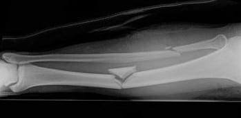
Butterflies
Butterfly fragments are large triangular fragments commonly seen in comminuted long bone fractures.
FFFF
Funtabulously Frivolous Friday Five
Dr Neil Long BMBS FACEM FRCEM FRCPC. Emergency Physician at Kelowna hospital, British Columbia. Loves the misery of alpine climbing and working in austere environments (namely tertiary trauma centres). Supporter of FOAMed, lifelong education and trying to find that elusive peak performance.


