


Gilula carpal arcs
Gilula carpal arcs (Gilula lines; three carpal arcs) are three smooth radiographic contours used to assess carpal alignment on a neutral posteroanterior (PA) wrist radiograph.
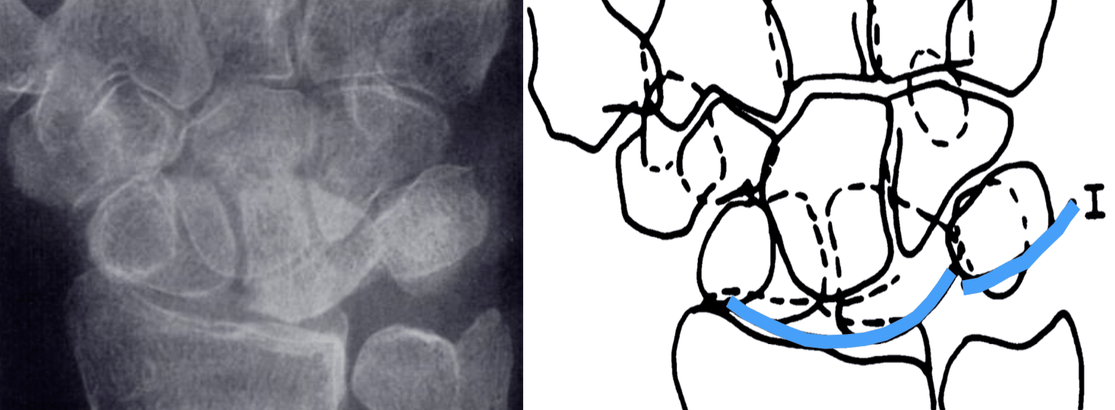
- Arc I follows the proximal convex margins of the scaphoid, lunate and triquetrum.
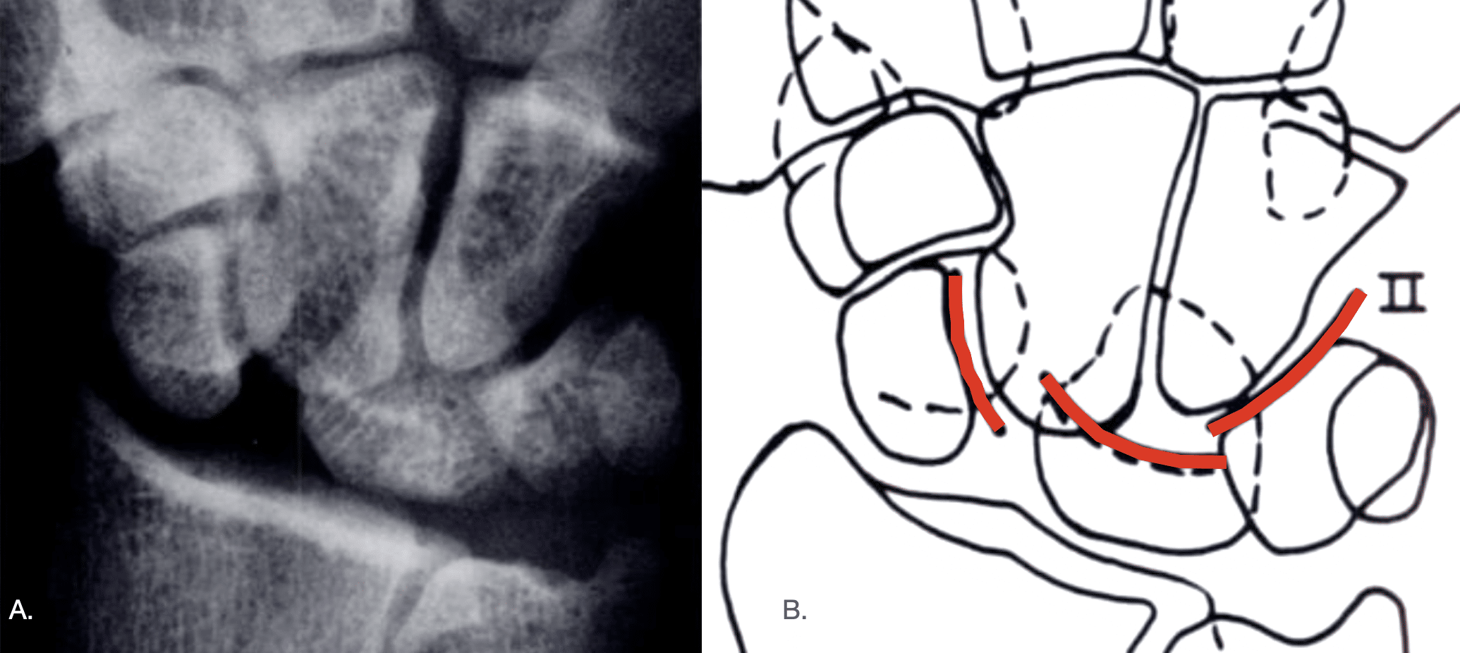
- Arc II follows the distal concave margins of the scaphoid, lunate and triquetrum.
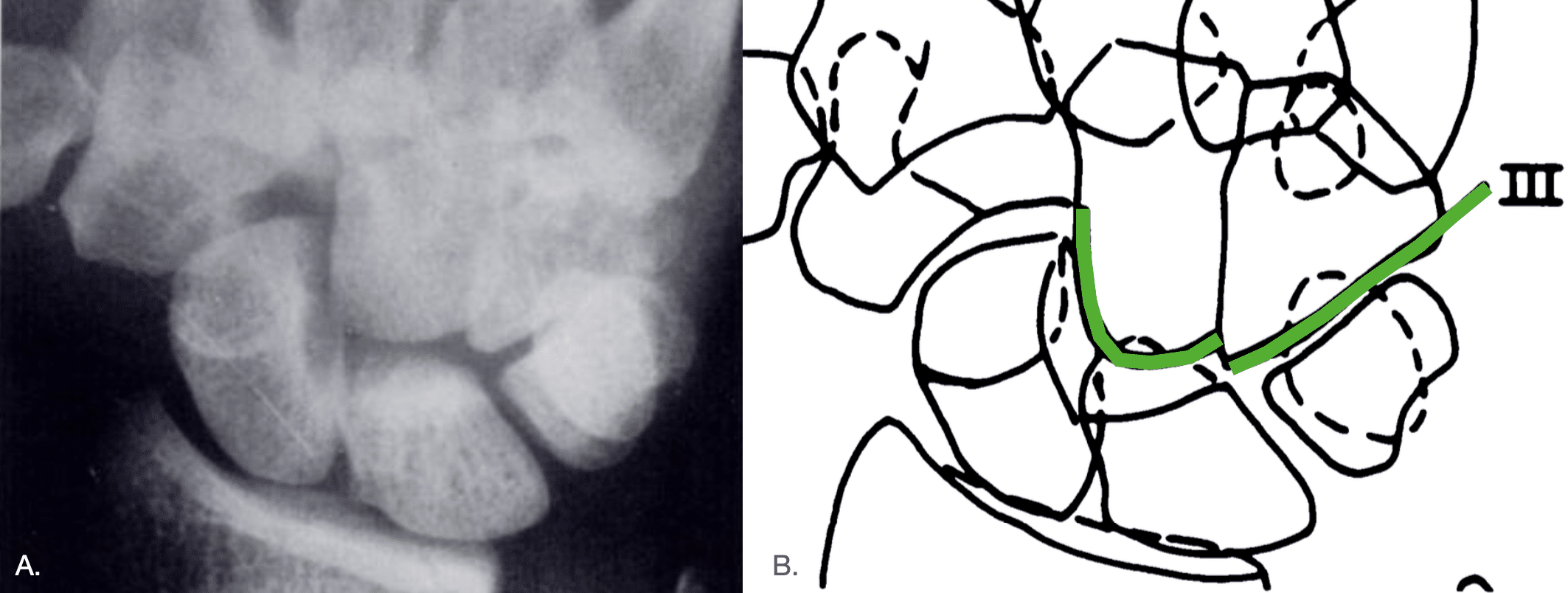
- Arc III follows the proximal convex margins of the capitate and hamate.
In a normally aligned neutral wrist the arcs form smooth, largely uninterrupted curves. A step-off, sharp angulation or break should prompt assessment for carpal malalignment, fracture or ligamentous injury at the site of disruption.
The arcs are a screening and localising tool rather than a stand-alone diagnostic test. Wrist positioning and normal anatomical variation may disrupt Arcs I and II without pathological instability.
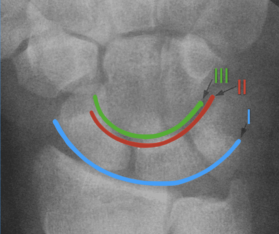
Arc I (blue) follows the proximal convex margins of the scaphoid, lunate and triquetrum;
Arc II (red) follows the distal concave margins of the same three bones; and
Arc III (green) follows the proximal convex margins of the capitate and hamate. The arcs should form smooth curves without a major step-off.
Fundamentals
Gilula described the arcs as part of a systematic method for interpreting carpal alignment rather than as isolated lines. A disrupted arc identifies where to look, while the joint-space, overlap and alignment abnormalities help determine what has happened.
Assessment begins with a properly positioned neutral PA wrist radiograph and asks:
- Are Arcs I, II and III smooth and continuous?
- Are the intercarpal joint spaces of similar width?
- Are opposing articular surfaces parallel where they are normally seen in profile?
- Is there abnormal overlap between normally articulating carpal bones?
- Is the shape or orientation of an individual carpal bone abnormal?
When to use
Gilula carpal arcs are useful as a rapid check for a subtle injury. They should be reviewed routinely on the PA radiograph of an injured wrist looking for
- subtle carpal malalignment;
- scapholunate or lunotriquetral injury;
- perilunate or lunate dislocation;
- carpal fracture-dislocation; or
- an associated carpal injury accompanying a more obvious distal radius or carpal fracture.
Interpreting disruption
A break in a Gilula arc is an alignment sign, rather than a diagnosis. The site of interruption localises the region requiring closer inspection. Depending on the associated findings, disruption may reflect ligamentous injury, fracture, subluxation or dislocation.
Multiple arcs may be abnormal in complex injuries, while an isolated disruption may provide the first clue to a comparatively subtle abnormality.
Normal variants and pitfalls
Apparent arc disruption is not always pathological. The the arcs should be assessed on a strictly neutral PA wrist radiograph and interpreted with the remaining carpal relationships.
- Wrist position: Arcs I and II commonly become discontinuous during radial or ulnar deviation. Arc III remained intact in every position.
- Triquetral morphology: A relatively short triquetrum may produce a normal step-off in Arc I at the lunotriquetral joint while Arc II remains smooth.
- Type II lunate: A medial hamate facet may alter the contour of the distal proximal-row surface and produce an apparent bilobed or interrupted Arc II.
Modern context
Gilula’s arcs remain a useful first-pass assessment of carpal alignment on plain radiographs. They do not exclude occult ligamentous or osseous injury when preserved, and an abnormal arc does not by itself establish the precise lesion.
Stress radiographs may demonstrate dynamic instability, while CT provides superior definition of complex fractures and fracture-dislocations. MRI, MR arthrography and wrist arthroscopy may be required when ligamentous injury remains suspected despite equivocal radiographs.
History of the carpal arcs
1896 – Thomas Hastie Bryce (1862–1946) published Certain Points in the Anatomy and Mechanism of the Wrist-Joint Reviewed in the Light of a Series of Röntgen Ray Photographs of the Living Hand one of the the earliest radiographic studies of wrist kinematics. His observations established the concept of proximal and distal carpal rows and differential movement between individual carpal bones.
1935 – Sir Roy Douglas Wright (1907–1990) recognised the circular radiographic contour formed by the proximal carpal row while studying wrist movement. In his Detailed Study of Movement of the Wrist Joint he described the first carpal row as forming an almost complete circular arc on the posteroanterior wrist radiograph (Gilula arc I). Gilula later acknowledged Wright’s observation as a precursor to his three-arc method.
1979 – Louis A. Gilula (1942–2014) reviewed more than 90 carpal fractures, fracture-dislocations and normal wrists. He described three radiographic carpal arcs on the PA wrist view as part of a systematic method for assessing carpal alignment. Discontinuity helped localise fracture, ligamentous disruption or carpal malalignment.
In the analysis of these cases, it became evident that on the posteroanterior radiograph, three fairly smooth radiographic arcs could be drawn to define normal carpal bone relationships (fig. 1 ). Break of any of these arcs strongly suggests abnormality at the site of the broken arc.
Arc I follows the main convex curvatures of the proximal surfaces of the scaphoid (navicular), lunate, and triquetrum carpal bones. Arc II outlines the distal concave curvatures of these same three bones. Arc III traces the main proximal curvatures of the capitate and hamate.
Gilula 1979
Arc I follows the proximal convex cortical margins of the scaphoid, lunate and triquetrum.
Arc II follows the distal concave cortical margins of the scaphoid, lunate and triquetrum.
In the figure below Arc II is interrupted at both the scapholunate and lunotriquetral joints. Associated widening and abnormal overlap of the carpal bones provided additional evidence that the proximal row was disrupted.
Arc III follows the major proximal curvatures of the capitate and hamate.
Unlike Arcs I and II, which outline the proximal carpal row, Arc III assesses alignment within the distal carpal row, particularly the relationship between the capitate and hamate. In the figure Arc III is interrupted at the capitohamate joint.
Disruption of the arcs
Gilula defined a broken arc as a localising clue within a broader system that examined:
- joint-space width and symmetry;
- parallelism of opposing articular surfaces;
- abnormal overlap of normally articulating surfaces;
- altered shape or orientation of individual carpal bones; and
- correlation between PA, oblique and lateral projections.
1996 – Wilfred C. G. Peh and Gilula examined Normal disruption of carpal arcs in 100 asymptomatic wrists. They found that Arcs I and II may normally become disrupted during radial or ulnar deviation, while they are usually continuous in a neutral wrist. Arc III remained intact in all positions studied. They concluded that wrist positioning must be considered before interpreting disruption of Arcs I or II as pathological.
1998 – Reynolds, Johnston and Friedman adapted Gilula’s arcs to a carpal stretch test for dynamic dissociative carpal instability. They examined six patients with arthroscopically proven proximal-row ligament injury but nondiagnostic routine radiographs. They found that traction produced measurable disruption of Arcs I and II and helped localise otherwise occult instability. The very small series demonstrated that the arcs could be used dynamically rather than solely on resting radiographs.
2008 – Kaewlai and colleagues analysed Multidetector CT of carpal injuries. CT provided rapid multiplanar and three-dimensional evaluation of carpal fractures and dislocations and could reveal fractures occult on radiographs. Gilula’s arcs remained a useful screening tool on the initial PA radiograph, but CT could further demonstrate complex three-dimensional osseous relationships.
2011 – Bong Cheol Kwon et al prospectively evaluated a modified carpal stretch test in 49 unstable intra-articular distal-radius fractures. They used disruption of Gilula Arc II as a marker of high-grade scapholunate ligament injury and arthroscopy as the reference standard. Mean sensitivity was 78%, specificity 72%, negative predictive value 87%, and overall accuracy 74%. They concluded that the test was more useful for excluding a major scapholunate tear than for confirming one.
2023 – Okoro et al reviewed Imaging Diagnosis and Management of Carpal Trauma and Instability. They suggested using Gilula’s arcs at the start of radiographic assessment of carpal alignment, along with scapholunate and capitolunate angles and other alignment criteria. CT, MRI and MR arthrography are then used to define fractures, ligamentous injury and complex instability that cannot be fully characterised by the arcs alone.
Associated Persons
- Thomas Hastie Bryce (1862–1946)
- Sir Roy Douglas Wright (1907–1990)
- Louis Arnold Gilula (1942-2014)
Alternative names
- Gilula three carpal arcs
- Gilula’s arcs, Gilula arcs
- Gilula’s lines, Gilula lines
References
Historical references
- Bryce TH. Certain Points in the Anatomy and Mechanism of the Wrist-Joint Reviewed in the Light of a Series of Röntgen Ray Photographs of the Living Hand. J Anat Physiol. 1896 Oct;31(Pt 1):59-79.
- Wright RD. A Detailed Study of Movement of the Wrist Joint. J Anat. 1935 Oct;70(Pt 1):137-142
- Gilula LA. Carpal injuries: analytic approach and case exercises. AJR Am J Roentgenol. 1979 Sep;133(3):503-17.
- Gilula LA. Wrist Trauma: Roentgenographic Analysis In: Traumatized Hand and Wrist: Radiographic and Anatomic Correlation. 1992: 221-239
- Peh WC, Gilula LA. Normal disruption of carpal arcs. J Hand Surg Am. 1996 Jul;21(4):561-6
Eponymous term review
- Reynolds RA, Johnston GH, Friedman L. The carpal stretch test. Can J Surg. 1998 Apr;41(2):119-26.
- Loredo RA, Sorge DG, Garcia G. Radiographic evaluation of the wrist: a vanishing art. Semin Roentgenol. 2005 Jul;40(3):248-89.
- Kaewlai R, Avery LL, Asrani AV, Abujudeh HH, Sacknoff R, Novelline RA. Multidetector CT of carpal injuries: anatomy, fractures, and fracture-dislocations. Radiographics. 2008 Oct;28(6):1771-84.
- Vezeridis PS, Yoshioka H, Han R, Blazar P. Ulnar-sided wrist pain. Part I: anatomy and physical examination. Skeletal Radiol. 2010 Aug;39(8):733-45.
- Kwon BC, Choi SJ, Song SY, Baek SH, Baek GH. Modified carpal stretch test as a screening test for detection of scapholunate interosseous ligament injuries associated with distal radial fractures. J Bone Joint Surg Am. 2011 May 4;93(9):855-62.
- Okoro CK, Skalski MR, Patel DB, White EA, Matcuk GR Jr. Imaging Diagnosis and Management of Carpal Trauma and Instability-An Illustrated Guide. Life (Basel). 2023 Jun 21;13(7):1426.
- Martínez-Carpio PA, Moreno Fructuoso M, Lleopart Rodríguez N, Bedoya Del Campillo Á. Trans-scapho-perilunate dislocation and Gilula’s arcs. Rev Esp Sanid Penit. 2025 Nov 18;27(3):135-137.
eponymictionary
the names behind the name

MBChB University of Manchester, PG Dip (SEM) University of Bath. Keen interest in Sport and Exercise Medicine. Currently working in emergency medicine in Perth, WA and intend to train as an ACSEP Registrar.
BA MA (Oxon) MBChB (Edin) FACEM FFSEM. Emergency physician, Sir Charles Gairdner Hospital. Passion for rugby, medical history, medical education, and asynchronous learning #FOAMed evangelist. Editor-in-Chief and Webmaster of Life in the Fast lane, LITFL | On Call 4e| Eponyms | Books | Horology |