


Louis Gilula

Louis Arnold Gilula (1942-2014) was an American radiologist
Gilula was a pioneer of musculoskeletal and wrist imaging. He spent 40 years at the Mallinckrodt Institute of Radiology (MIR), Washington University School of Medicine, where he became professor of radiology, orthopaedic surgery, plastic and reconstructive surgery.
Gilula is best remembered for his systematic approach to carpal radiography with the Gilula carpal arcs, described in 1979. He also contributed to early interventional radiology, image-guided treatment of spinal pain, vertebroplasty and other percutaneous procedures.
Biographical Timeline
- Born on October 21, 1942 in Lubbock, Texas son of Dr Adolph and Dora Gilula
- 1967 – Graduated MD from the University of Illinois School of Medicine, Chicago. Completed internship at San Francisco General Hospital.
- 1968–1970 – Began radiology training in St Louis but was drafted after six weeks of residency. Served as a Captain in the United States Army, with postgraduate training at DeWitt Army Hospital, Fort Belvoir, Virginia.
- 1970–1973 – Returned to St Louis and completed radiology residency at St Louis City Hospital (Max C. Starkloff Memorial Hospital).
- 1973 – Joined the Mallinckrodt Institute of Radiology (MIR), Washington University School of Medicine. Over the following 40 years he rose to professor of radiology, orthopaedic surgery, and plastic and reconstructive surgery, and headed the musculoskeletal section for 24 years.
- 1979 – Published Carpal injuries: analytic approach and case exercises, introducing the three radiographic carpal arcs that subsequently became known as Gilula arcs.
- Co-founded the International Wrist Investigators Workshop and was its only radiologist member. With hand surgeon Julio Taleisnik, helped guide the Workshop for more than two decades as an international forum for wrist research.
- 1992 – Consolidated his work on midcarpal arthrography, carpal instability, ulnar translation, wrist fractures and degenerative disease in his textbook The Traumatized Hand and Wrist: Radiographic and Anatomic Correlation.
- 2008–2010 – Recognised repeatedly for teaching at MIR. Distinguished Teacher of the Year (2008/2009) and Teacher of the Year in 2010.
- 2013 – Retired from Washington University in October after approximately 40 years at Mallinckrodt. The department conference room was dedicated as the Louis A. Gilula, MD Conference Room in recognition of his contribution to education and training.
- Died July 2, 2014 aged 71, following pancreatic cancer in Ladue, St. Louis County
Medical Eponyms
Gilula carpal arcs (1979)
Gilula’s three carpal arcs are assessed on a posteroanterior wrist X-ray. They represent the smooth cortical contours formed by the proximal and distal carpal rows. Gilula developed the system after reviewing over 90 carpal fractures and fracture-dislocations between 1973 and 1978.
In the normal wrist, small indentations may occur where adjacent bones meet, but the overall curvature should remain smooth. Gilula emphasised that interruption of an arc should prompt careful examination of the corresponding joint or bone.
In the analysis of these cases, it became evident that on the posteroanterior radiograph, three fairly smooth radiographic arcs could be drawn to define normal carpal bone relationships (fig. 1 ). Break of any of these arcs strongly suggests abnormality at the site of the broken arc.
Arc I follows the main convex curvatures of the proximal surfaces of the scaphoid (navicular), lunate, and triquetrum carpal bones. Arc II outlines the distal concave curvatures of these same three bones. Arc III traces the main proximal curvatures of the capitate and hamate.
Gilula 1979
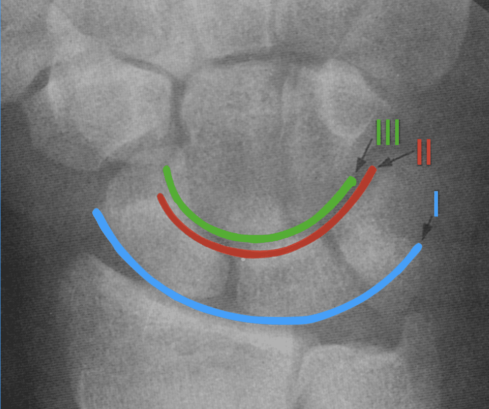
Arc I (blue) follows the proximal convex margins of the scaphoid, lunate and triquetrum;
Arc II (red) follows the distal concave margins of the same three bones; and
Arc III (green) follows the proximal convex margins of the capitate and hamate. The arcs should form smooth curves without a major step-off.
1996 – Wilfred C. G. Peh and Gilula examined Normal disruption of carpal arcs in 100 asymptomatic wrists. They found that Arcs I and II may normally become disrupted during radial or ulnar deviation, while they are usually continuous in a neutral wrist. Arc III remained intact in all positions studied. They concluded that wrist positioning must be considered before interpreting disruption of Arcs I or II as pathological.
Key Medical Contributions
Interventional radiology and treatment of spinal pain
Gilula was an early contributor to interventional radiology. His work included angiography of haemodialysis arteriovenous fistulas, catheter-directed thrombolytic therapy, therapeutic spinal injections and other image-guided percutaneous procedures. He became an early adopter and investigator of vertebroplasty, and published on its indications, contraindications and technique.
His clinical practice included vertebroplasty, kyphoplasty, cementoplasty, radiofrequency treatment of painful osseous lesions and image-guided management of chronic spinal pain. Washington University later described him as a pioneer who offered therapeutic spinal injections before such treatment became commonplace.
Teaching and scholarship
Gilula regarded teaching as a central part of his career. During his 40 years at Mallinckrodt he taught generations of radiology residents and fellows, receiving repeated institutional teaching awards and, on retirement, having a departmental conference room named in his honour. He authored more than 300 peer-reviewed articles, two textbooks, and numerous chapters, reviews and case reports.
Major Publications
- Gilula LA, Staple TW. Minature atlas of total hip prostheses. Radiol Clin North Am. 1975 Apr;13(1):21-44.
- Ford LT, Gilula LA, Murphy WA, Gado M. Analysis of gas in vacuum lumbar disc. AJR Am J Roentgenol. 1977 Jun;128(6):1056-7.
- Gilula LA, Murphy WA, Tailor CC, Patel RB. Computed tomography of the osseous pelvis. Radiology. 1979 Jul;132(1):107-14.
- Gilula LA. Carpal injuries: analytic approach and case exercises. AJR Am J Roentgenol. 1979 Sep;133(3):503-17. [Gilula carpal arcs]
- Conway WF, Destouet JM, Gilula LA, Bellinghausen HW, Weeks PM. The carpal boss: an overview of radiographic evaluation. Radiology. 1985 Jul;156(1):29-31.
- Gilula LA, Reinus WR, Totty WG. Midcarpal wrist arthrography. AJR Am J Roentgenol. 1986 Mar;146(3):645-7.
- Gilula LA. Wrist Trauma: Roentgenographic Analysis In: Traumatized Hand and Wrist: Radiographic and Anatomic Correlation. 1992: 221-239
- Peh WC, Gilula LA. Normal disruption of carpal arcs. J Hand Surg Am. 1996 Jul;21(4):561-6
- Goldfarb CA, Yin Y, Gilula LA, Fisher AJ, Boyer MI. Wrist fractures: what the clinician wants to know. Radiology. 2001 Apr;219(1):11-28.
- Gilula LA, Lander P. Management of spinal pain with imaging-guided injection. Radiographics. 2003 Jan-Feb;23(1):189-90; author reply 190-1.
- Peh WC, Gilula LA. Percutaneous vertebroplasty: indications, contraindications, and technique. Br J Radiol. 2003 Jan;76(901):69-75.
- Wollstein R, Wei C, Bilonick RA, Gilula LA. The radiographic measurement of ulnar translation. J Hand Surg Eur Vol. 2009 Jun;34(3):384-7
- White L, Clavijo J, Gilula LA, Wollstein R. Classification system for isolated arthritis of the scaphotrapeziotrapezoidal joint. Scand J Plast Reconstr Surg Hand Surg. 2010 Apr;44(2):112-7
References
Biography
- Purdy MC. Obituary: Louis Gilula, professor of radiology, 71. Wash U The Source. July 3, 2014
- Slutsky DJ. Louis Gilula, MD-A Tribute. J Wrist Surg. 2014 Aug;3(3):161.
- Rubin D. Louis A. Gilula, MD. Radiology. 2015 Jan;274(1):308.
- Dr Louis Arnold Gilula. Obituary
Eponymous terms
- Wright RD. A Detailed Study of Movement of the Wrist Joint. J Anat. 1935 Oct;70(Pt 1):137-142
- Reynolds RA, Johnston GH, Friedman L. The carpal stretch test. Can J Surg. 1998 Apr;41(2):119-26.
- Loredo RA, Sorge DG, Garcia G. Radiographic evaluation of the wrist: a vanishing art. Semin Roentgenol. 2005 Jul;40(3):248-89.
- Kaewlai R, Avery LL, Asrani AV, Abujudeh HH, Sacknoff R, Novelline RA. Multidetector CT of carpal injuries: anatomy, fractures, and fracture-dislocations. Radiographics. 2008 Oct;28(6):1771-84.
- Vezeridis PS, Yoshioka H, Han R, Blazar P. Ulnar-sided wrist pain. Part I: anatomy and physical examination. Skeletal Radiol. 2010 Aug;39(8):733-45.
- Kwon BC, Choi SJ, Song SY, Baek SH, Baek GH. Modified carpal stretch test as a screening test for detection of scapholunate interosseous ligament injuries associated with distal radial fractures. J Bone Joint Surg Am. 2011 May 4;93(9):855-62.
- Okoro CK, Skalski MR, Patel DB, White EA, Matcuk GR Jr. Imaging Diagnosis and Management of Carpal Trauma and Instability-An Illustrated Guide. Life (Basel). 2023 Jun 21;13(7):1426.
- Knipe H. Gilula three carpal arcs. Radiopaedia
Eponym
the person behind the name

MBChB University of Manchester, PG Dip (SEM) University of Bath. Keen interest in Sport and Exercise Medicine. Currently working in emergency medicine in Perth, WA and intend to train as an ACSEP Registrar.

BA MA (Oxon) MBChB (Edin) FACEM FFSEM. Emergency physician, Sir Charles Gairdner Hospital. Passion for rugby, medical history, medical education, and asynchronous learning #FOAMed evangelist. Editor-in-Chief and Webmaster of Life in the Fast lane, LITFL | On Call 4e| Eponyms | Books | Horology |