![]()
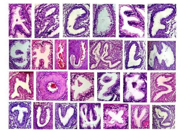
Glandular Alphabetum
Trawling the internet for useful resources on histology…late on a Friday night (= nerd), I happened across this amazing alphabetum made from glands!
Dr. Ma. Ivy Clemente took these microscope photos of glandular structures from Fibroadenoma (benign breast tumor) and Nodular Prostatic Hyperplasia (non-cancerous prostate growth) cases
iHeartGuts.com
This inspired me to keep looking, and I found a sample chapter (Chapter 7 Nervous System) of the much anticipated 2nd edition of Functional histology by Dr Jeffrey B. Kerr – Some amazing histologic images that come closer to what is seen down the microscope than any other source I have found. Amazing images, beautifully presented, well worth a look.
Pathology must be really boring sometimes…all that purple and pink and cold laboratory environment. So who can blame them for being excited when they chance upon a pareidolic experience, a Wolkenkuckucksheim if you like where the pathologist finally has the chance to say ‘Bad news: you have a tumor. Good news: it’s really cute!‘

BA MA (Oxon) MBChB (Edin) FACEM FFSEM. Emergency physician, Sir Charles Gairdner Hospital. Passion for rugby; medical history; medical education; and asynchronous learning #FOAMed evangelist. Co-founder and CTO of Life in the Fast lane | On Call: Principles and Protocol 4e| Eponyms | Books |