ICE 006: Knee injury X-ray
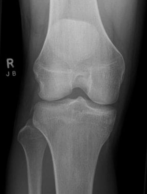
A 24 year old man is brought into the ED with an injury suffered playing touch rugby. He tried to stop and turn suddenly just as another player collided with him. He felt immediate right knee pain and has only been able to hobble a few steps since.
Questions:
- What abnormality can be seen on his AP X-ray?
- Explain the significance of this finding
- What treatment is required?
Reveal the ICE answer
The xray findings are subtle and consist of a fine bone fragment (about 10mm long and up to 4mm wide) seen at the border of the upper part of the lateral tibial plateau. This is a so called “Segond” fracture
The unimpressive radiographic findings belie the severity of the underlying knee injury. The mechanism of the Segond fracture is a combination of bowing and twisting (technically a combination of varus stress and internal tibial rotation) of the knee . The bone fragment seen is an avulsion fracture from the lateral ligament complex but the real significance is that there are usually also injuries to the anterior cruciate and medial meniscus. So it represents a major knee joint disruption with potential for significant instability.
The most appropriate management of meniscal and cruciate injuries is controversial and will vary in elite versus recreational athletes. Nevertheless the presence of a Segond fracture makes chronic instability and pain likely so these patients should have an early orthopaedic assessment. An MRI would be indicated if surgery is planned. In the interim, standard care will include RICE, analgesia, splint and a short period of non weight bearing.
References
- Eponymictionary – Paul Ferdinand Segond (1851-1912)
- Eponymictionary – Segond fracture

ICE CASES
Ian’s clinical emergencies
emergency physician keen on medical education and cycling

