![]()
Segond fracture
Description
Segond fracture: Avulsion fracture (small) of the lateral surface of the lateral tibial condyle. Usually results from excessive internal rotation and varus stress resulting in increased tension on the lateral capsular ligament of the knee joint. Frequently (75-100% of cases) associated with disruption of the anterior cruciate ligament (ACL).
In the majority of cases a segond avulsion fracture is associated with detachment of the capsular portion of the lateral collateral ligament and tears of the anterior cruciate ligament. Additional injury to menisci and other supporting ligaments.
Anterior cruciate ligament injury is most commonly associated with valgus stress, however, the Segond fracture usually occurs as a result of internal rotation and varus stress. Most commonly associated with sporting injuries especially soccer, skiing, basketball and baseball.
Clinical implication of the Segond fracture
- Segond fracture associated with increased probability of other soft tissue such as ACL rupture; posterolateral horn injury; MCL rupture; or meniscal injury
- Segond fracture present in 3–7% of all ACL ruptures, although there is an ACL injury present in 75–100% of the Segond fractures.
Imaging
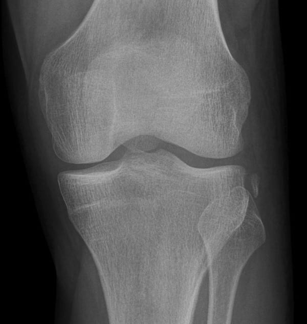
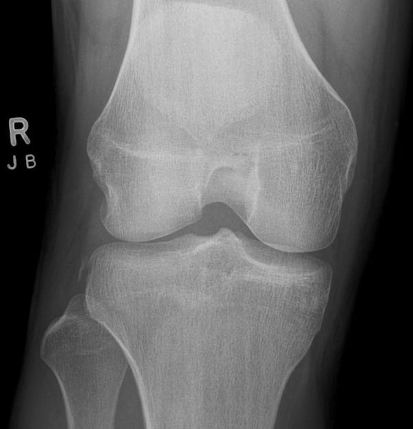
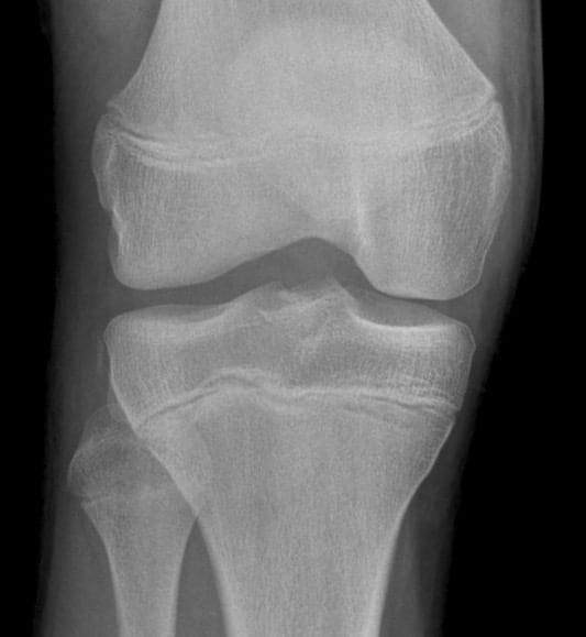
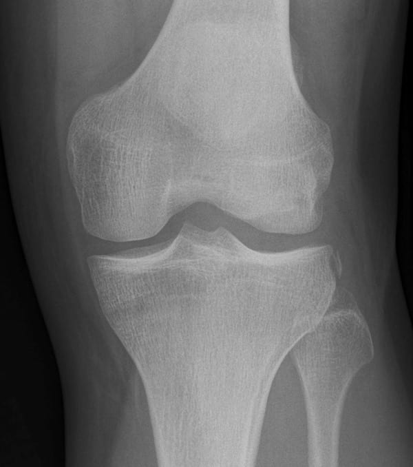
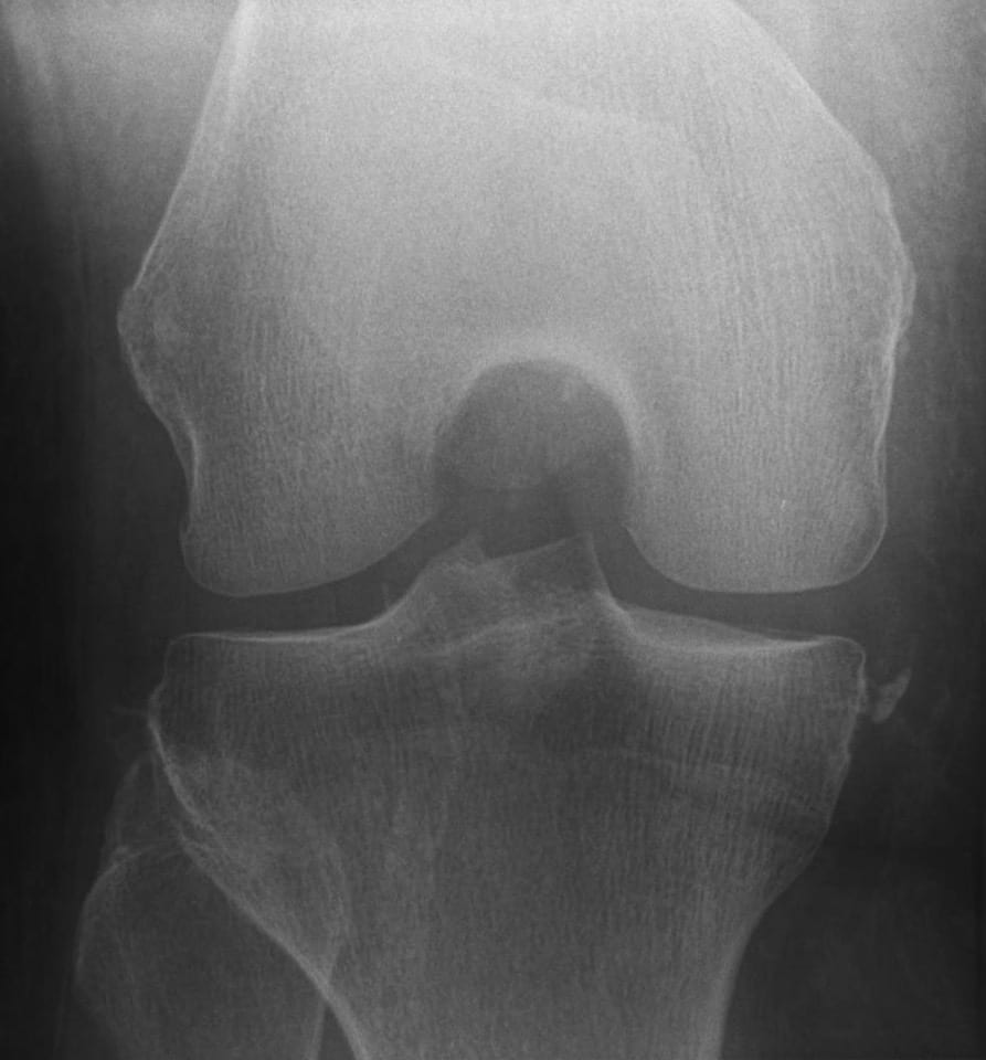
X-Ray: The classical appearance of a Segond fracture is that of an elliptic or curvilinear bone fragment parallel to the lateral aspect of the tibial plateau. Best seen on the anteroposterior view of the knee.
MRI: Segond fractures are often associated with internal knee injuries and MRI is essential. Associated injuries include:
- ACL tear: most common associated injury; occurring in 75-100% of cases
- Medial or lateral meniscal tear; 66-75% of cases; posterior horn most commonly affected.
- Long head of biceps femoris avulsion
- Fibular collateral ligament avulsion


History of the Segond fracture
1845 – Amédée Bonnet published the first cadaveric studies for the mechanism of knee ligament injuries. Bonnet concluded (Traité des maladies des articulations: 1845; II: 188) that following forced movements of extension and laterality:
- On well-formed individuals, these forced movements never cause fractures of the bones, but tearing of the ligaments, muscular ruptures
- No permanent dislocation occurs, if the two portions of the lower limb are mobile, and thus contribute to the production of forced movement
- On subjects of advanced age, on children and on adults of poor constitution, the fracture of the articular extremities is the almost inevitable consequence of these kinds of violence.
- These fractures, which can simulate dislocations, are difficult to heal due to the meshing of the surfaces and the crushing of the bones.
1879 – Paul Segond acknowledged and repeated the work of Bonnet.
Repeating the experiments of Bonnet, we were determined to produce anatomic lesions of a knee sprain, and to then, by performing meticulous dissection, look for a tear or any lesion that would be sufficient in its extent, nature and location to produce bleeding inside the joint.
He demonstrated (in 17 of 38 experiments with cadavers) that internal rotation of the tibia with the knee in flexion results in tension on the lateral joint capsule of the knee at its midpoint where a pearly, fibrous, resistant band of tissue (the lateral capsular ligament (LCL)), produces an avulsion fracture of the lateral tibia.
The lateral ligaments do not “always remove their point of insertion to the femur” as Bonnet says. Its assertion is true only for the internal lateral ligament, the lower insertion point of which indeed remains always intact; but the inferior insertion of the external lateral ligament may very well give way. We observed it in one case.
Segond 1879
Segond described the small bony avulsion on the lateral tibial plateau and commented that:
It is never Gerdy’s tubercle that gives way, but the portion of bone immediately behind it…the lesion is pathognomonic of torsion of the knee in internal rotation and slight flexion of the lower leg and is associated with rupture of the anterior cruciate ligament

Segond went on to describe signs and symptoms of anterior cruciate rupture:
…strong articular pain, frequent accompanying pop, rapid joint effusion and abnormal anterior-posterior movement of the knee on clinical examinations
1936 – Henry Milch first reported this fracture on radiographs of acutely injured knees in three patients.
This avulsion is caused by tension on the iiotibial band at its insertion into the area behind Gerdy’s tubercle. It is usually acquired by injury, with the knee in the partly flexed position, but it may also be produced with the knee in the fully flexed position, if the heel is held medially to the axis of the femur during the internal rotation of the leg

In reviewing the literature, the author could find no clinical information on this interesting lesion. By accident, a reference to the experimental work of Segond was noted. Though no clinical cases are cited in this work, it appears that, in studying the effects of rotation, Segond accurately described the pathology of the type of cortical avulsion fracture which has been mentioned.
Milch 1936
Associated Persons
- Pierre Nicolas Gerdy (1797-1856) [Gerdy tubercle]
- Amédée Bonnet (1809-1858) [Original cadaveric experiments]
- Paul Ferdinand Segond (1851-1912)
- Henry Milch (1895-1964) [First use of the Segond eponym]
Segond fracture examples
Note: Reverse Segond
Reverse Segond fracture: Avulsion of the deep fibers of the medial collateral ligament. Usually located along the medial proximal tibia adjacent to the articular surface. Most commonly associated with valgus stress and external rotation of the knee [Opposite of Segond fracture which involves the avulsion fragment of the lateral proximal tibia following knee varus stress and internal rotation]
References
Original articles
- Bonnet A. Traité des maladies des articulations. Paris: Baillière. 1845 Tome Second, Atlas. [Segond 1845; II: 188]
- Segond P. Recherches cliniques et expérimentales sur les épanchements sanguins du genou par entorse. Progrès Médical 1879; 16: 297–299, 319–321, 340–341
- Milch H. Cortical avulsion fracture of the lateral tibial condyle. J Bone Joint Surg 1936; 18(1): 159-164.
Review articles
- Dietz GW, Wilcox DM, Montgomery JB. Segond tibial condyle fracture: lateral capsular ligament avulsion. Radiology. 1986; 159(2): 467-9.
- Goldman AB, Pavlov H, Rubenstein D. The Segond fracture of the proximal tibia: a small avulsion that reflects major ligamentous damage. AJR Am J Roentgenol. 1988; 151(6): 1163-7.
- Davis DS, Post WR. Segond Fracture: Lateral Capsular Ligament Avulsion. JOSPT. 1997; 25(2): 102-106
- Campos JC et al. Pathogenesis of the Segond fracture: anatomic and MR imaging evidence of an iliotibial tract or anterior oblique band avulsion. Radiology. 2001; 219(2): 381-6
- Gottsegen CJ, Eyer BA, White EA, Learch TJ, Forrester D. Avulsion fractures of the knee: imaging findings and clinical significance. Radiographics. 2008; 28(6): 1755-70.
- Valkering KP, Breederveld RS. Segond fracture. J Am Coll Surg 2009; 208: 646
- Schindler OS. Surgery for anterior cruciate ligament deficiency: a historical perspective. Knee Surg Sports Traumatol Arthrosc. 2012; 20(1): 5-47.
- Wharton R et al. Segond fracture in an adult is not pathognomonic for ACL injury. Knee Surg Sports Traumatol Arthrosc 2015; 23:1925–1928
- Somford MP et al. Biographical background and origin of common eponymous terms in orthopedic surgery: anatomy and fractures in knee surgery. Eur J Orthop Surg Traumatol. 2018 Jan;28(1):79-84
- Murgier J et al. The Origin of the Knee Anterolateral Ligament Discovery: A Translation of Segond’s Original Work With Commentary. Arthroscopy. 2019; 35(2): 684-690
- Rasuli B. Segond Fracture. Radiopaedia
- Cadogan M. Segond fracture. Eponym A Day. Instagram
[cite]
BA MA (Oxon) MBChB (Edin) FACEM FFSEM. Emergency physician, Sir Charles Gairdner Hospital. Passion for rugby; medical history; medical education; and asynchronous learning #FOAMed evangelist. Co-founder and CTO of Life in the Fast lane | On Call: Principles and Protocol 4e| Eponyms | Books |