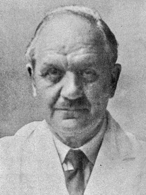
Ivor Lewis

Ivor Lewis (1895–1982) was a Welsh surgeon
Lewis was a demon for work and a hard taskmaster who made as heavy demands on himself as on his medical and nursing teams.
On January 10, 1946 Lewis delivered the Hunterian Lecture on the surgical treatment of carcinoma of the oesophagus at the RoyalCollege of Surgeons of England. This procedure is known eponymously as the Ivor Lewis procedure
He dearly loved his native Wales and its culture and was a fluent Welsh speaker. He became
member of the White Order of the Gorsedd of Bards (bardic name Ifor o Wynfe) and the National Eisteddfod and he was an elder of the Calvinistic Methodist Church at Cefn Meiriadog. Staunch supporter of the Welsh Surgical Society, (President 1959 and 1960. Long-standing member of the Welsh Regional Hospital Board contributing to the wider development of health services in his homeland.
Biography
- Born on October 27, 1895 at Llanddeusant, Carmarthenshire the only child of farmer Lewis Lewis
- 1915-1918 Pre-clinical studies at University College, Cardiff
- 1919-1921 Clinical training at University College Hospital, London. [MRCS LRCP 1920; MB BS 1921; Lister Gold Medal in surgery 1921]
- 1922-1930 Resident surgical officer at Lewisham Hospital [MD 1924; MS 1930]
- 1930 – Surgeon and medical director at Plymouth City Hospital. Fostered the practice whereby patients could be visited by their relatives on a daily basis, by no means a common practice in those days
- 1933-1951 Consultant surgeon and medical director, North Middlesex Hospital
- 1939 – Performed the first pulmonary embolectomy operation in Great Britain
- 1948 – FRCS by election
- 1951 – Moved back to Wales to allow his four children a Welsh education. Surgeon Royal Alexandra Hospital in Rhyl; and the chest hospitals at Abergeleand and Llangwyfan
- 1970 – member of the White Order of the Gorsedd of Bards (bardic name Ifor o Wynfe), admitted at the National Eisteddfod in Ammanford, Carmarthenshire
- 1977 – DSc honoris causa, at the University of Wales
- Died on September 11, 1982 at St Asaph, Wales
- 1983 – Ivor Lewis Memorial Lecture established at the Postgraduate Education Centre at Glan Clwyd Hospital
Medical Eponyms
Ivor Lewis procedure
An upper gastrointestinal tract operation performed for mid and distal oesophageal pathology, usually oesophageal cancer.
In 1946 described a two-staged oesophagectomy, including a laparotomy followed by a right-sided thoracotomy, and an immediate intrathoracic gastroesophageal anastomosis.
The new approach advocated immediate rather than delayed reconstruction and involved two standardized incisions, the Ivor Lewis procedure gained immediate popularity and is now the most commonly utilized approach for esophageal resection worldwide.
An account is given of a new operation of resection with immediate anastomosis for cancer in the middle third. After first mobilizing the stomach by laparotomy, a right transpleural oesophagectomy is performed, the stomach being brought up through the dilated hiatus, fixed at any required level in the right pleura, and an end-to-side oesophagogastrostomy performed.


Major Publications
- Lewis I. Trendelenburg’s operation for pulmonary embolism a successful case. 1939; 233(6036): 1037-1041
- Lewis I. Hospital visiting. Lancet 1943; 241(6237): 348.
- Lewis I. Carcinoma of the Œsophagus. Radical Resection with Œsophago-gastrostomy for a Midthoracic Growth by a Right Transpleural Approach. Proc R Soc Med. 1945 Jul;38(9):483-4.
- Lewis I. The surgical treatment of carcinoma of the oesophagus; with special reference to a new operation for growths of the middle third. Br J Surg. 1946 Jul;34:18-31. [Being the substance of a Hunterian Lecture at the Royal College of Surgeons, London, Jan. 10, 1946]
References
Biography
- Franklin RH. Ivor Lewis Lecture, 1975. The advancing frontiers of oesophageal surgery. Ann R Coll Surg Engl. 1977 Jul;59(4):284-7.
- Obituary. Ivor Lewis. The Lancet. 1982; 320(8301): 779
- Obituary. Ivor Lewis. Br Med J (Clin Res Ed) 1982;285:982
- Buddug Owen. Ifor o Wynfe: Cofiant byr i Ifor Lewis y Llawfeddyg. 1991
- Morris-Stiff G, Hughes LE. Ivor Lewis (1895-1982) – Welsh pioneer of the right-sided approach to the oesophagus. Dig Surg. 2003;20(6):546-52; discussion 552-3.
- Lewis, Ivor (1895-1982), consultant surgeon. Y Bywgraffiader Cymraig
- Biography: Lewis, Ivor (1895-1982). Plarr’s Lives of the Fellows Online. Royal College of Surgeons of England.
Eponymous terms
- Turner GG. Excision of the thoracic oesophagus for carcinoma: with construction of an extra-thoracic gullet. Lancet. 1933; 222(5754):1315-1316
- Lerut T, Wiesel O. History of esophagectomy for cancer of the esophagus and the gastroesophageal junction. Ann Transl Med. 2021 May;9(10):897.
- Qaraqe TM, Low DE. Ivor Lewis Esophagectomy. Atlas of Esophageal Surgery 2022: 125-138
- Rucker AJ, D’Amico TA, Pappas TN. Ivor Lewis Esophagectomy and the Care of Humphrey Bogart’s Midesophageal Cancer. Ann Thorac Surg. 2024 Aug;118(2):510-516.
Eponym
the person behind the name
