![]()
Lung abscess
A previously healthy 22 year old man with a 4 week history. He describes the cough, some mild shortness of breath on exertion, purulent sputum, fevers, night sweats and weight loss.
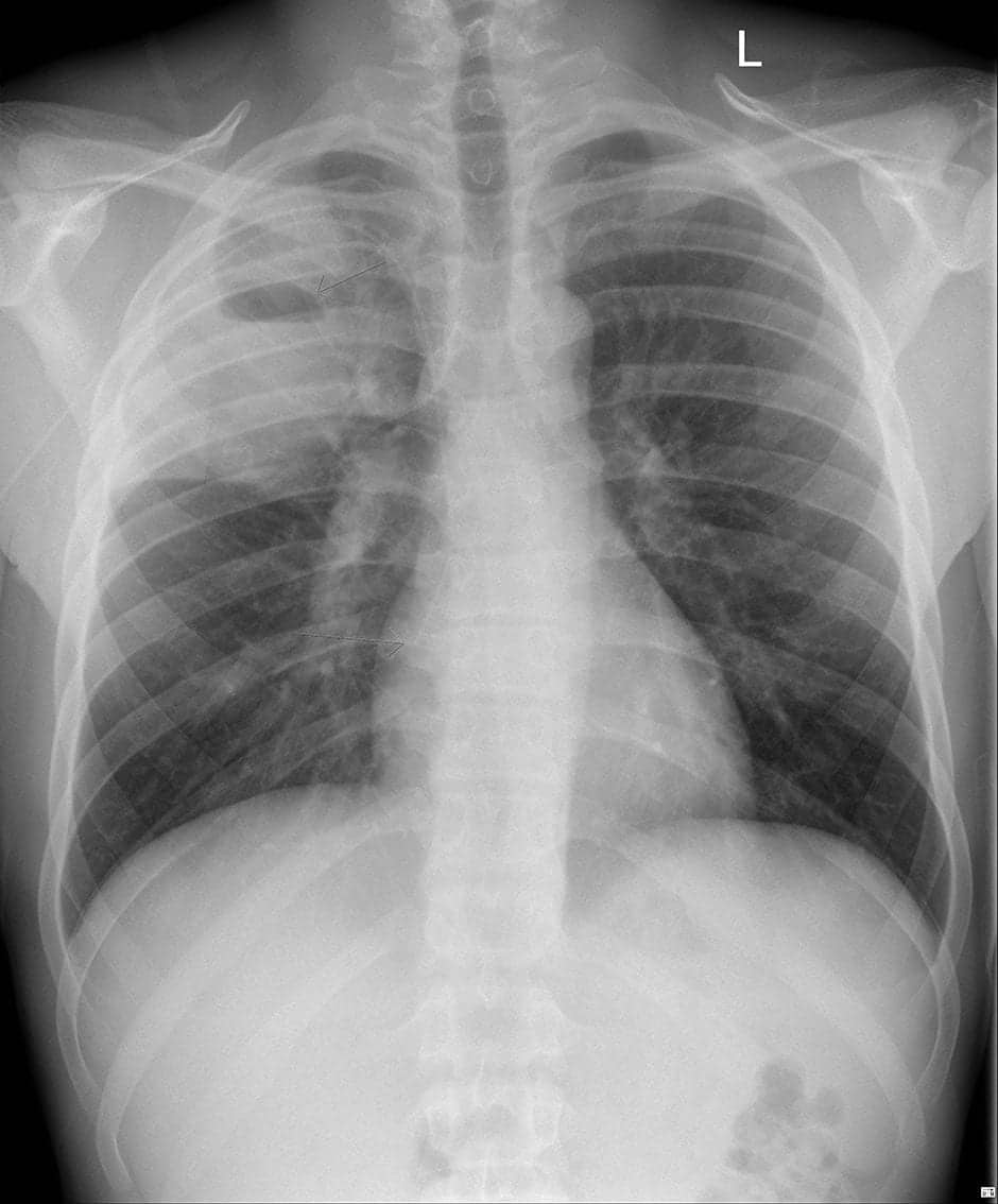
His chest x-ray shows right upper lobe consolidation with an air fluid level
You perform an ultrasound to characterize the consolidated area. Describe your findings.
Ultrasound View 1
Reveal Answer
The anterior approach to the right upper lobe only reveals normal lung. You explore the apex of the lung posteriorly.
The typical ultrasound pattern of consolidation with hepatisation is absent. There has been destruction of lung parenchyma which has been replaced by fluid and multiple air locules. This is a complex lung abscess.
The respiratory team arrange a CT for further characterization.
Show Images and Explanation
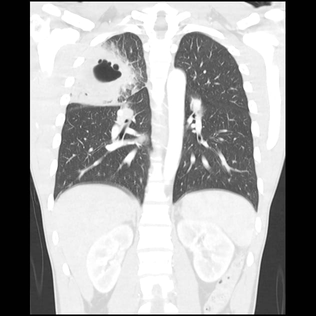
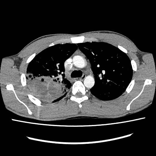
- The CT shows the complex RUL abscess with loculations and multiple air locules.
- The differential for a cavitating lung lesion is large. In a young otherwise healthy Australian adult, preceding symptoms typical for pneumonia and no significant risk factors or exposures the most common aetiology would be Streptococcus pneumoniae.
Related Clinical Cases
- LITFL Ultrasound library
- LITFL Top 100 ultrasound cases
- LUNG ultrasound cases
- LUNG ultrasound modules

ULTRASOUND LIBRARY
Clinical Cases

An Emergency physician based in Perth, Western Australia. Professionally my passion lies in integrating advanced diagnostic and procedural ultrasound into clinical assessment and management of the undifferentiated patient. Sharing hard fought knowledge with innovative educational techniques to ensure knowledge translation and dissemination is my goal. Family, wild coastlines, native forests, and tinkering in the shed fills the rest of my contented time. | SonoCPD | Ultrasound library | Top 100 | @thesonocave |