![]()
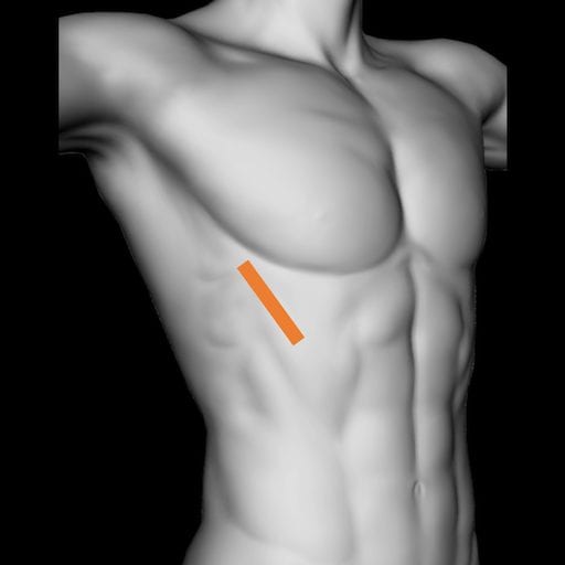
Lung ultrasound: Lung Point
In the presence of a pneumothorax the visceral and parietal pleural surfaces are separated. The point at which these two surfaces meet is known as the lung point
Finding a typical lung point confirms that the absence of sliding is due to a pneumothorax.The position of the lung point gives some information on the size and location of the pneumothorax.
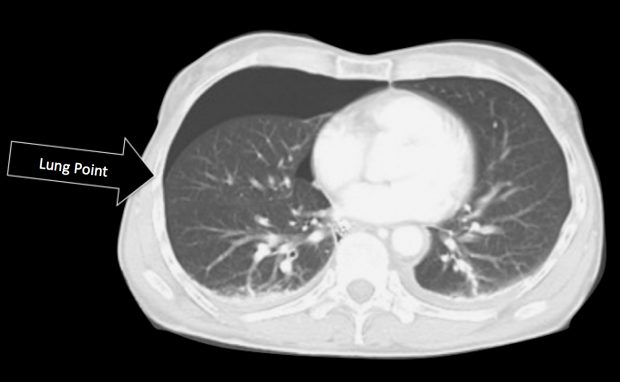
A typical lung point confirming pneumothorax
Ultrasound appearance of lung point explained
Ultrasound appearance of lung point explained
- A typical lung point is sensitive and specific for pneumothorax.
- In the presence of a pneumothorax the visceral pleura falls away from the parietal pleura. The point at which the two pleural surfaces touch is called the lung point. Unless there are pleural adhesions this point is dynamic and moves with the respiratory and cardiac cycle.
- The position of the lung point depends on the size of the pneumothorax
- Where the two pleural surfaces are touching ultrasound features of normal lung are seen.
- Where the two pleural surfaces are separated the ultrasound features of pneumothorax are evident.
- Note: The lung point is sometimes referred to as the contact point.
Related Clinical Cases
- LITFL Ultrasound library
- LITFL Top 100 ultrasound cases
- LUNG ultrasound WORKED CASES
- LUNG ultrasound MODULES

ULTRASOUND LIBRARY
Modules

An Emergency physician based in Perth, Western Australia. Professionally my passion lies in integrating advanced diagnostic and procedural ultrasound into clinical assessment and management of the undifferentiated patient. Sharing hard fought knowledge with innovative educational techniques to ensure knowledge translation and dissemination is my goal. Family, wild coastlines, native forests, and tinkering in the shed fills the rest of my contented time. | SonoCPD | Ultrasound library | Top 100 | @thesonocave |