![]()
No Radial Pulse
aka Cardiovascular Curveball 012
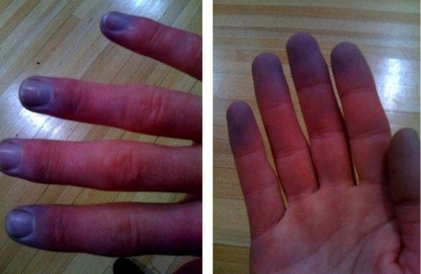
Priority 1 Call: 68 year-old female who has central chest pain 2/10 after the administration of aspirin and GTN, and a past history of angina.Worryingly, the patient’s finger tips have turned blue, and the paramedics are unable palpate a radial pulse…
On arrival in the ED, the patient is sitting up, alert and orientated, and is in no distress or pain anymore. You look at her hands:
Q1. What is the most likely diagnosis?
Answer and interpretation
Raynaud’s phenomenon
Raynaud’s phenomenon is episodic vasospasm of the peripheral arteries. It characteristically causes pallor followed by cyanosis and redness with pain and sometimes paraesthesia (white then blue then red colour change.
On rare occasions it can lead to ulceration of the fingers and toes (and, in some cases, the ears or nose).
Q2. How is this condition classified? What are the distinguishing features?
Answer and interpretation
Raynaud’s affects women more than men, in a 9:1 ratio, and most are affected under the age of 25 years. Around 20% of people that are diagnosed with primary Raynaud’s phenomenon go onto to develop another autoimmune disease such as systemic lupus erythematous or rheumatoid arthritis.Raynaud’s may be primary or secondary.
- Primary Raynaud’s is idiopathic (no known cause), and usually follows a benign course,
- Secondary Raynaud’s are caused by underlying disease.
Primary Raynaud’s
- Episodic colour changes evoked by cold or emotional stimuli
- Patient is not known to have an underlying condition that causes Raynaud’s
- No digital scars or gangrene
- Normal ESR
- Negative antinuclear antibodies
- Normal chest X-ray
- Even nail-fold after capillaroscopy
Secondary Raynaud’s
- Episodic colour changes evoked by cold or emotions
- Patient is known to have a condition that causes Raynaud’s
- Digital scars or gangrene
- Abnormal ESR
- Positive antinuclear antibodies
- Abnormal chest X-rays
- Uneven nail-fold after capillaroscopy
Q3. What are the causes and risk factors for this condition?
Answer and interpretation
Raynaud’s phenomenon may be a result of any of the following:
- Scleroderma
- Systemic lupus erythematosis
- Rheumatoid arthritis
- Sjögren’s syndrome
- Carpal tunnel syndrome
- Repetitive trauma
- Smoking
- Drugs, such as beta blockers and ergotamine
- Chemical exposures, including exposure to vinyl chloride
Raynaud’s phenomenon has also been linked to hyperthyroidism and pulmonary hypertension.
Q4. What are the signs and symptoms of this condition?
Answer and interpretation
The affected body part turns:
- White (pallor) as the small blood vessels constrict then
- Bluish (cyanosis) occurs when there is a slight relaxation of arterial spasm resulting in a slight flow of blood into the dilated capillary bed, where it rapidly dissipates, and finally back to
- Red (normal) and the vasospasm decreases, and the blod vessels become dilated again allowing blood flow to return
The following questions will help in making the diagnosis:
- Are your fingers unusually sensitive to cold?
- Do your fingers change colour when they exposed to cold temperatures?
- Do they turn white, blue, or both?
Q5. What management do these patients require in the emergency department?
Answer and interpretation
Emergency department care is centered on reassurance, symptomatic relief (generally with non-drug therapy), and appropriate followed up with either primary care provider or specialist rheumatologist.
Acute management options:
- Reassurance
- Provide simple analgesia, e.g. paracetamol, NSAIDs, codeine
- Immerse affected parts in warm water
- Gently massage and rub affected areas until warm
- Use relaxation techniques if condition is stressed induced
- Encourage patients to remain indoors on cold days
- Promote smoking cessation
The evidence base for preventative measures is limited. Medications such as nifedipine and prazosin may be used to reduce the frequency and severity of attacks.
References:
- Pope, J. (2008). Raynaud’s Phenomenon (primary). Clinical Evidence. [PMC2907991]
- Summers, A. (2005). From white, to blue, to red: Raynaud’s Phenomenon. Emergency Nurse. 2005 Nov;13(7):18-20 [PMID: 16318066]
- Wigley,F. (2002). Raynaud’s Phenomenon. NEJM. Sep 26;347(13):1001-8 [PMID: 12324557]
CLINICAL CASES
Cardiovascular Curveball
Emergency nurse with ultra-keen interest in the realms of toxicology, sepsis, eLearning and the management of critical care in the Emergency Department | LinkedIn |