
![]()
Pneumonia Case 001
This patient presented with right upper quadrant pain and a fever. The clinical suspicion was cholecystitis.
What does this clip show?
Reveal Answer
- This RUQ view shows a small area of basal consolidation.
- The normally reflective aerated lung in the posterior costophrenic angle is lost, and replaced by a tiny pleural effusion surrounding consolidated lung.
- The “spine sign” is seen and is characteristic of basal pathology. The posterior chest wall is usually obscured by air in normal lung above the diaphragm. In this case the spine can be seen and followed cranially above the diaphragm. This is because ultrasound can penetrate the fluid and solid lung, rather than being reflected away completely.
A second view is taken higher and more laterally over the area of interest in the lower chest wall. This allows direct visualization of the lung rather than looking through liver and diaphragm.
What does the ultrasound show?
View 2
Reveal Answer
- A small region of basal consolidation is confirmed, with a surrounding pleural effusion.
- The patient’s clinical presentation was consistent with a basal pneumonia and the ultrasound features confirmed this.
- They include consolidated lung, air bronchograms, an irregular “shredded” border between the consolidated and aerated lung, with B-lines at the interface, and an associated small pleural effusion.
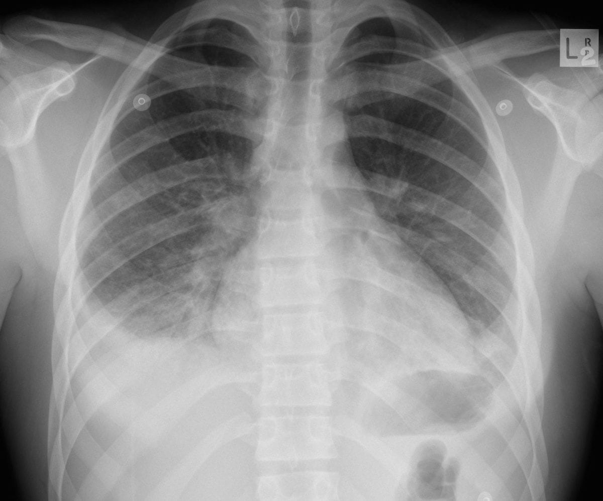
Chest X-ray performed after the ultrasound confirms the findings of right lower lobe consolidation.
What does this histological sample demonstrate?
Reveal Answer
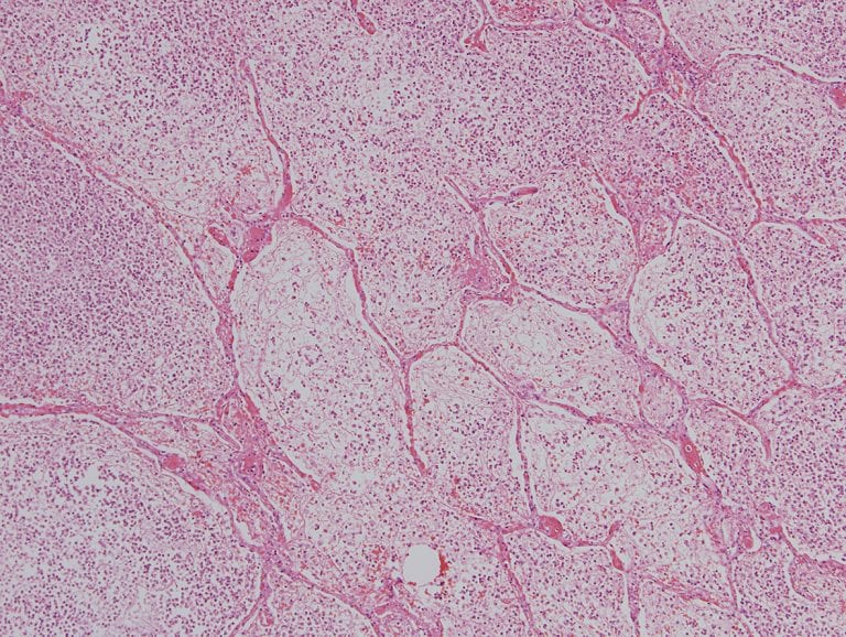
- This slide is a histological section of bronchopneumonia.
- Air spaces are completely filled with inflammatory cells and exudate.
- This explains why in pneumonia, ultrasound can penetrate the lung parenchyma rather than being reflected away by air.Where air remains within bronchi the characteristic “sonographic air bronchogram” is seen.
Related Clinical Cases
- LITFL Ultrasound library
- LITFL Top 100 ultrasound cases
- LUNG ultrasound cases
- LUNG ultrasound modules

ULTRASOUND LIBRARY
Clinical Cases

An Emergency physician based in Perth, Western Australia. Professionally my passion lies in integrating advanced diagnostic and procedural ultrasound into clinical assessment and management of the undifferentiated patient. Sharing hard fought knowledge with innovative educational techniques to ensure knowledge translation and dissemination is my goal. Family, wild coastlines, native forests, and tinkering in the shed fills the rest of my contented time. | SonoCPD | Ultrasound library | Top 100 | @thesonocave |