![]()
Pneumothorax Case 5
A 36 year old woman presents with sharp left sided chest pain. What do these clips of her left chest show?
View 2
View 3
Reveal Answer
- The first clip shows the typical features of pneumothorax.
- On the left of the image the sternum is seen, then the internal thoracic vessels.
- The echogenic line deep to these is the pleural surface and there is a complete lack of sliding. The vertical movement of the pleural surface is chest wall movement with respiration.
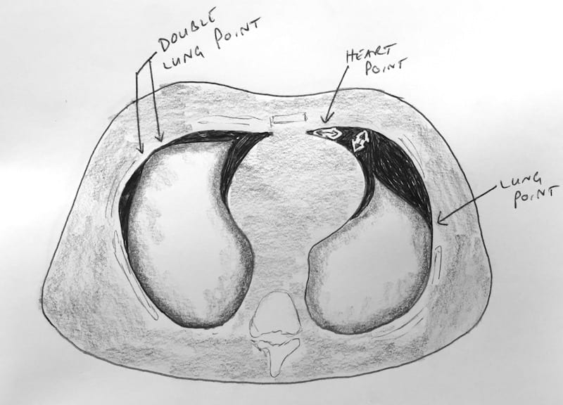
- The second clip shows a double contact point, where lung abuts the pleural surface with free air on either side.
- The final clip shows a typical lung point with sliding lung on the right and pneumothorax on the left.
Explanation of the various lung points.
The patient’s chest x-ray. The small apical pneumothorax is very subtle.

Related Clinical Cases
- LITFL Ultrasound library
- LITFL Top 100 ultrasound cases
- LUNG ultrasound cases
- LUNG ultrasound modules

ULTRASOUND LIBRARY
Clinical Cases

An Emergency physician based in Perth, Western Australia. Professionally my passion lies in integrating advanced diagnostic and procedural ultrasound into clinical assessment and management of the undifferentiated patient. Sharing hard fought knowledge with innovative educational techniques to ensure knowledge translation and dissemination is my goal. Family, wild coastlines, native forests, and tinkering in the shed fills the rest of my contented time. | SonoCPD | Ultrasound library | Top 100 | @thesonocave |