
![]()
Reed-Sternberg cells
Description
Reed-Sternberg cells are large, abnormal lymphocytes (a type of white blood cell) that may contain more than one nucleus. These cells are found in people with Hodgkin lymphoma.
Hodgkin and Reed-Sternberg cells are the hallmark cells in Hodgkin’s disease. The are the tumor cells of Hodgkin lymphoma and are usually termed Hodgkin cells when mononucleated and Reed-Sternberg cells when multinucleated.
The cells are derived from (pre-apoptotic) germinal center B cells, but have have lost their B cell identity and show aberrant activation of multiple signalling pathways.
The cells are named after Thomas Hodgkin for his description of a lymphoid lesion in 1832; and Carl Sternberg (1898) and Dorothy Reed (1902) for their later histologic description of the cells. There is dispute of the eponymic recognition of Reed-Sternberg cells, and to whom credit for their recognition lies. Willis (1948) argued that Greenfield, Sutton and Coupland had “clearly depicted” the cells in 1878. However, it is accepted that none of the earlier descriptions of this particular cell are fully admissible, whereas Sternberg and Reed’s description come close.
History of the Reed-Sternberg cell
1872 – Theodor Langhans (1839-1915) was one of the first to describe the histopathologic features of Hodgkin’s disease and to recognize the presence of multinucleated giant cells.
1878 – Symposium, chaired by Wilks, on the subject of lymphadenoma and leukaemia at the Pathological Society of London. This included several significant contributions referring to the large multinucleated tumour cells of Hodgkin’s disease. William Smith Greenfield (1846 – 1919) provided specimens illustrative of the pathology of lymphadenoma and leucocythemia. He described five cases, all with similar microscopic appearances:
…there was some general enlargement of the glands, the fibrous stroma appeared coarser, and there were a large number of multinucleated cells adherent to the trabeculae…These multinucleated cells, containing from four to eight or twelve nuclei, were often collected in dusters in parts of the gland especially where the fibrous change was progressing.
Greenfield 1878
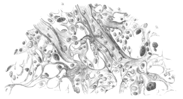
1898 – Carl Sternberg (1872-1935) at the University of Vienna published 15 cases of Hodgkin’s disease with illustrations. He described the microscopic appearances of the lymph glands, the multinucleated giant cells; the mononuclear variant; mitotic figures; and distinguished the giant cells from the Langhans giant cells found in tuberculosis.
Original
English
Sie sind scharf begrenzt, zeigen stellenweise peripher noch Reste von Anhäufungen lymphatischer Zellen, meist aber ist auch diese wie die übrigen Partien umgewandelt in ein stellenweise mehr zellarmes, lockeres, fibröses Gewebe, indem sich Spindelzellen und vereinzelte Lymphocyten und Leukocyten finden, ferner grössere, protoplasmareiche Zellen mit grossen, dunkelgefärbten Kernen, die nicht so selten mehrfach zu zweien und . dreien oder auch gelappt erscheinen. Vereinzelte solche Zellen besitzen eine grössere Anzahl (bis zu fünf und sechs) Kernen. An manchen sind die Kerne auffallend gross, rund und enthalten sich mit Eosin tingierende Kernkörperchen, oder, was seltener der Fall ist, die Kerne sind blass gefärbt und enthalten einen oder zwei sich mit Eosin färbende Nucleolen.
They are sharply defined, show in places peripherally residues of accumulations of lymphatic cells but most of the tissue is altered to a focally hypocellular, loose fibrous tissue in which spindle cells and single lymphocytes and leukocytes occur, also larger protoplasm-rich cells with large dark-stained nuclei, which not rarely appear multiple, up to 2 or 3 or also lobulated. Individual giant cells may have up to five or six nuclei. In some, the nuclei are strikingly big, round…and contain one or two eosinophilic nucleoli.
There are also large pale eosinophilic protoplasmic bodies with a pale eosinophilic, large
nucleus, structures corresponding to the large cells.
Sternberg essentially provided an accurate description of the microscopic pathology and illustrated the giant cells. However, as 8 of the 15 cases demonstrated tuberculosis in one or more organ, Sternberg concluded that Hodgkin’s disease was a chronic inflammatory process secondary to the tubercle bacillus, a belief he held until his death in 1935.
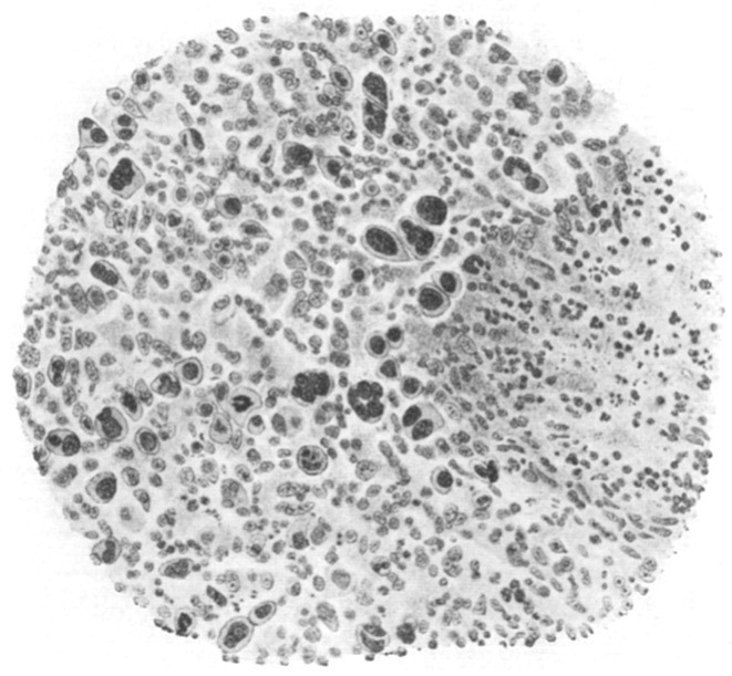
1901-1902 During her pathology fellowship at Johns Hopkins, Dorothy Reed (1874-1964) reviewed surgical specimens and autopsy material of cases that were considered to be Hodgkin’s disease. She found 8 cases which closely resembled each other clinically and histologically.
Reed firmly asserted the nature of Hodgkin’s disease:
We should limit the term Hodgkin’s disease to designate a clinical and pathological entity, the main features of which are painless progressive glandular enlargements, usually starting in the cervical region, without the blood changes of leukemia.
With respect to Hodgkin’s disease and tuberculosis:
The outcome of all this discussion seems to be that tubercle bacilli may be present in a malignant lymphomatous process, without any primary etiological connection between the two, and also that there may be advanced tuberculosis present in a case of malignant lymphoma without the presence of tubercle bacilli in the gland
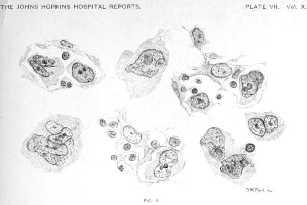
Fig 4. The so-called endothelial cells with their branching protoplasmic processes in different stages of proliferation. Transition from uninuclear giant cells, originating from such endothelial cells, to large multinuclear giant cells are represented, showing the peculiar vesicular nuclei, prominent nucleoli, and protoplasmic processes
Reed accurately described and illustrated the cells characteristic of Hodgkin’s disease:
The nucleus is always large in proportion to the size of the cell. It may be single or multiple. If single, it is usually round. Bean-shaped and irregularly indented nuclei are common. If multiple, the nuclei may be arranged peripherally in the cell, or heaped in the center. Eight or ten nuclei have been seen in a single cell. The chromatin network is prominent in these nuclei, and one or more large nucleoli are always present. The nucleoli are usually oval, but they may be of any shape. The nucleoli always take a contrasting stain to the nucleus in the double stains, they have an affinity for acid dyes. No definite mitotic figures were ever seen in these cells. Direct division was frequently observed. The protoplasm is usually homogeneous, and stains well. It may appear granular, show vacuolization, or contain fat or pigment granules. Cells having bizarre and irregular nuclei are found in the oldest growths. These giant cells, so far as our observation reaches, are peculiar to this growth, and are of great assistance in diagnosis
Reed. 1902
1902 – Sir Frederick William Andrewes (1859-1932) independently published an illustrated description of the histopathology of Hodgkin’s disease having reviewed more than 20 cases over 3 years. He described the large multinucleated cells of Hodgkin’s disease (Reed-Sternberg cells) as well as nodular sclerosing Hodgkin’s disease. [Discussion on lymphadenoma in its relation to tuberculosis. Transactions of the Pathological Society of London]
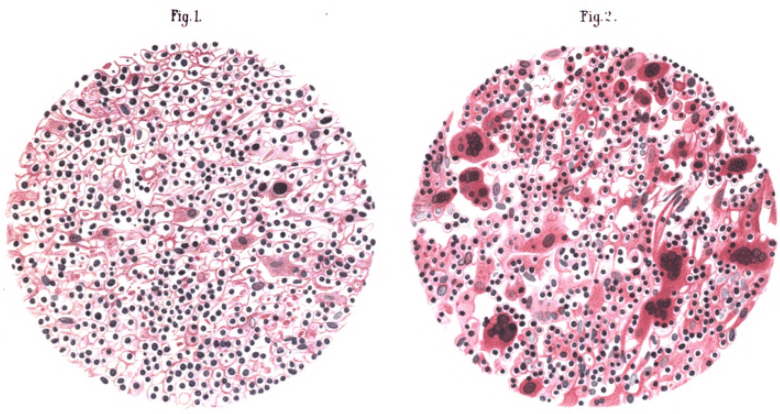
- Fig 1. Section of an axillary lymphatic gland removed from a man aged 38. Emphasizes the pleocellularity of process, together with the giant cells
- Fig 2. Section of a cervical lymphatic gland removed from a man aged 26 affected with lymphadenoma. Large numbers and great size of the ‘lymphadenoma cells’. These are for the most part multinucleate and deeply stained, but do not resemble the giant – cells of tubercle .

- Fig 3. Section of a cervical gland removed from a woman aged 32. Illustration of probable nodular sclerosing Hodgkin’s disease
- Fig 4. Section of a lymphatic gland from the axilla of a boy aged 11. Characteristic multinucleate giant cells
1916 – Reed’s name was not initially linked to the giant multinucleated cell of Hodgkin’s disease. However William George MacCallum (1874-1944) mentions Dorothy Reed five times in a 6-page chapter on Hodgkin’s disease in his Text-book on Pathology. This irreversibly associated Reed’s name with the cell in various forms including Sternberg-Reed; Reed-Sternberg; and Dorothy Reed cell.
Associated Persons
- Thomas Hodgkin (1798-1866)
- Theodor Langhans (1839-1915)
- William Smith Greenfield (1846-1919)
- Carl Sternberg (1872-1935)
- Dorothy Mabel Reed Mendenhall (1874-1964)
- Frederick William Andrewes (1859-1932)
Alternative names
- Dorothy Reed cell
- Reed-Sternberg cell
- Sternberg-Reed Cell
References
Original articles
- Langhans T. Das maligne Lymphosarkom (Pseudoleukämie). Archiv für pathologische Anatomie und Physiologie und für klinische Medicin, 1872; 54: 509–537.
- Greenfield WS. Specimens illustrative of the pathology of lymphadenoma and leucocythemia. Transactions of the Pathological Society of London, 1878; 29: 272–304
- Coupland S. Intestines and abdominal lymph glands from a case of lymphadenoma. Transactions of the Pathological Society of London, 1878; 29: 363-368
- Sternberg C. Über eine eigenartige unter dem Bilde der Pseudoleukämie verlaufende Tuberculose des lymphatischen Apparates. Zeitschrift für Heilkunde, 1898;1 9: 21-90.
- Reed DM. On the pathological changes in Hodgkin’s disease, with especial reference to its relation to tuberculosis. Johns Hopkins Hospital Report 1902; 10: 113-196
- Andrewes FW. Discussion on lymphadenoma in its relation to tuberculosis. Transactions of the Pathological Society of London, 1902; 53: 305-314
- MacCallum WG. Hodgkin’s disease or lymphogranulomatosis. In: A text-book of pathology. 1916: 791-796
Review articles
- Willis RA. The tumours of lymphoid tissue. In: Pathology Of Tumours. 1948: 74-75
- Rather LJ. Who discovered the pathognomonic giant cell of Hodgkin’s disease? Bull N Y Acad Med. 1972; 48(7): 943-950.
- Strum SB. The natural history, histopathology, staging, and mode of spread of Hodgkin’s disease. Series haematologica. 1973; 6(1): 20-115.
- Dawson PJ. The original illustrations of Hodgkin’s disease. Ann Diagn Pathol. 1999;3(6):386-393.
- Kaplan HS. Historical aspects. In: Hodgkin’s disease. 2e. 1980: 8-10
- Küppers R, Hansmann ML. The Hodgkin and Reed/Sternberg cell. Int J Biochem Cell Biol. 2005;37(3):511-517.
- Hong L. Thomas Hodgkin (1798 – 1866). LITFL 2020
- Nassar S. What did Dorothy Reed See? Hektoen International 2020
[cite]
eponymictionary
the names behind the name

Graduated from Cardiff Medical School in 2017 with MBBCh and BSc in Psychology and Medicine. Currently working as a doctor in the emergency department at Sir Charles Gairdner Hospital in Perth, Australia.
BA MA (Oxon) MBChB (Edin) FACEM FFSEM. Emergency physician, Sir Charles Gairdner Hospital. Passion for rugby; medical history; medical education; and asynchronous learning #FOAMed evangelist. Co-founder and CTO of Life in the Fast lane | On Call: Principles and Protocol 4e| Eponyms | Books |
