![]()
Dorothy Reed
Dorothy Mabel Reed Mendenhall (1874-1964) was an American physician, pediatrician and public health specialist.
In 1902, Reed, as the first female pathology fellow at Johns Hopkins, published research which determined that Hodgkin’s disease was not a form of tuberculosis. She demonstrated that the growth had a definite cellular structure of which a peculiar form of giant cell was the prominent feature.
She is remembered by the Dorothy Reed Mendenhall Scholarship Fund at Johns Hopkins University started in 1957 “to help a deserving woman medical student.”, and the Sabin-Reed Hall at Smith College. She is eponymously remembered for her description of Reed-Sternberg cells (1902).
Biography
- Born Dorothy Mabel Reed on September 22, 1874 in Columbus, Ohio.
- 1891-1895 Graduated B.L. from Smith College in Northampton, Massachusetts
- 1896 – Enrolled at John Hopkins Medical School
- 1898 – Along with fellow medical student Margaret Long, became the first women employed by a United States naval hospital assisting in the operating room and bacteriologic laboratories at the Brooklyn Navy Yard Hospital
- 1900 – MD, Graduated in medicine from the Johns Hopkins University School of Medicine and received a medical internship at Johns Hopkins Hospital under Sir William Osler (1849-1919) alongside Florence Sabin (1871-1953)
- 1901 – Fellowship in pathology under the direction of William Welch (1850-1934). Reed taught bacteriology, assisted at autopsies, and undertook research on Hodgkin’s disease, then believed to be a form of tuberculosis.
- 1902 – Reed published her historic paper on Hodgkin’s disease that subsequently gave her name to the Reed-Sternberg cell. However, following her research Reed approached Welch about applying for a staff position at Johns Hopkins. She records in her memoirs that he responded that Welch responded:
…no woman had ever held a teaching position in the school and that he [Welch] knew there would be great opposition to it. Suddenly, as I saw what I had to face in acceptance of injustice and in being overlooked, I knew that I could not take it. And I told Dr. Welch that if I could not look forward to a definite teaching position even after several years of apprenticeship, that I could not stay…
Reed, unpublished memoirs
- 1902 – Left Johns Hopkins and pathology to commence an interim residency at the New York Infirmary for Women and Children
- 1903 – First resident physician at Babies Hospital in New York City. Following the sudden death of her sister, Dorothy took on the financial responsibilities of looking after her sisters three children.
- 1906 – Married physicist Charles Elwood Mendenhall (1872-1935), whom she had known since childhood, and moved to Madison where he was professor of physics at the University of Wisconsin. Dorothy and Charles had 4 children: Margaret (1907) died of birth complications; Richard (1908) died in a fall aged 18 months; Thomas Corwin (1910), who later served as president of Smith College; and John Talcott (1912), who became a professor of surgery at the University of Wisconsin in Madison.
- 1914 – Lecturer in the Department of Home Economics at the University of Wisconsin
- 1915 – Organized the first infant welfare clinic in Wisconsin under the aegis of the Attic Angel Association and the Visiting Nurse Association and remained Chairman until 1936. Four more centers were opened.
- 1916-1935 Faculty member of the University of Wisconsin. Developed a correspondence course, the Nutrition Series for Mothers, for the Department of Agriculture (1918). Prepared six chapters for famous Children’s Bureau publication Child care and child welfare (1921)
- 1917-1936 Medical officer for the United States Children’s Bureau. Represented the bureau at the International Child Welfare Conference (1919) and helped to design the minimal standards for health centers.
- 1926 – Investigated infant and maternal mortality rates in Denmark compared to America, concluding that the higher death rates in America were due to unnecessary medical intervention.
- 1934 – Honorary Doctor of Science, Smith College for her contributions to infant welfare in Madison which had the lowest infant mortality in the nation for a city of its size (30/1,000 live births)
- 1935 – Following the death of her husband, Dorothy withdrew from active work. She travelled extensively and settled in Connecticut.
- Died July 31, 1964 in Chester, Connecticut at the age of 89
Medical Eponyms
Reed-Sternberg cells (1902)
In 1832, Thomas Hodgkin (1798-1866), reported the “morbid appearance of the absorbent glands and spleen” in patients with Hodgkins disease. This is later accepted to be due to the appearance of Reed-Sternberg calls.
Reed-Sternberg cells (variably termed “Sternberg,” “Dorothy Reed,” or “Sternberg-Reed” cells), are widely accepted to be pathognomonic of Hodgkin’s disease. They are precisely defined histologically to distinguish them from other large cell types.
Sternberg’s description of these cells was published in 1898, and Reed’s in 1902. There is much debate as to whether these cells had in fact been defined prior to this. There is dispute of the eponymic recognition of Reed-Sternberg cells, and to whom credit for their recognition lies. Willis (1948) argued that Greenfield, Sutton and Coupland had “clearly depicted” the cells in 1878.
It is accepted that none of the earlier descriptions of this particular cell are fully admissible, whereas Sternberg and Reed’s description come close.
1898 Sternberg published a paper describing “peculiar tuberculosis of the lymphatic apparatus running under the pattern of pseudoleukemia”
An abundance of peculiar mononuclear or multinuclear cells rich in protoplasm and with large, round to oval, or multiform, indented, lobate deeply stained nuclei in which nucleoli well-preserved mitotic figures were often visible.
[These cells] Often gave the tissue a peculiarly characteristic appearance…and strongly resembled tumour cells.
Sternberg 1898: 29
1901-1902 During her pathology fellowship at Johns Hopkins, Dorothy Reed reviewed surgical specimens and autopsy material of cases that were considered to be Hodgkin’s disease. She found 8 cases which closely resembled each other clinically and histologically.
Reed firmly asserted the nature of Hodgkin’s disease:
We should limit the term Hodgkin’s disease to designate a clinical and pathological entity, the main features of which are painless progressive glandular enlargements, usually starting in the cervical region, without the blood changes of leukemia.
With respect to Hodgkin’s disease and tuberculosis:
The outcome of all this discussion seems to be that tubercle bacilli may be present in a malignant lymphomatous process, without any primary etiological connection between the two, and also that there may be advanced tuberculosis present in a case of malignant lymphoma without the presence of tubercle bacilli in the gland
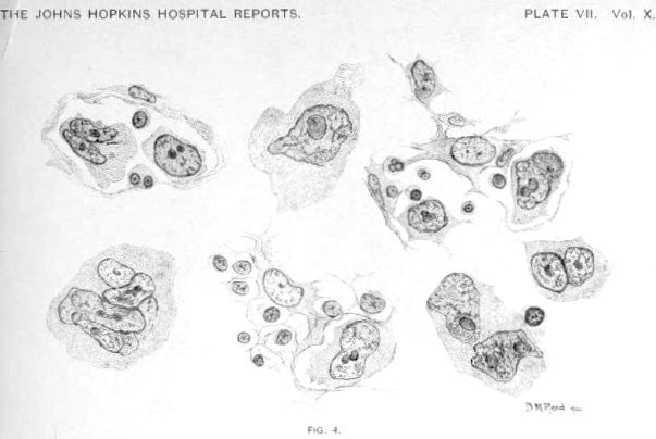
Dorothy Reed 1902: 196 Plate VII Fig 4
Fig 4. The so-called endothelial cells with their branching protoplasmic processes in different stages of proliferation. Transition from uninuclear giant cells, originating from such endothelial cells, to large multinuclear giant cells are represented, showing the peculiar vesicular nuclei, prominent nucleoli, and protoplasmic processes
Reed accurately described and illustrated the cells characteristic of Hodgkin’s disease:
The nucleus is always large in proportion to the size of the cell. It may be single or multiple. If single, it is usually round. Bean-shaped and irregularly indented nuclei are common. If multiple, the nuclei may be arranged peripherally in the cell, or heaped in the center. Eight or ten nuclei have been seen in a single cell. The chromatin network is prominent in these nuclei, and one or more large nucleoli are always present. The nucleoli are usually oval, but they may be of any shape. The nucleoli always take a contrasting stain to the nucleus in the double stains, they have an affinity for acid dyes. No definite mitotic figures were ever seen in these cells. Direct division was frequently observed. The protoplasm is usually homogeneous, and stains well. It may appear granular, show vacuolization, or contain fat or pigment granules. Cells having bizarre and irregular nuclei are found in the oldest growths. These giant cells, so far as our observation reaches, are peculiar to this growth, and are of great assistance in diagnosis.
Reed. 1902
1916 – Reed’s name was not initially linked to the giant multinucleated cell of Hodgkin’s disease. However William George MacCallum (1874-1944) mentions Dorothy Reed five times in a 6-page chapter on Hodgkin’s disease in his Text-book on Pathology. This irreversibly associated Reed’s name with the cell in various forms including Sternberg-Reed; Reed-Sternberg; and Dorothy Reed cell.
Major Publications
- Reed DM: The bacillus pseudo-tuberculosis murium; its streptothrix forms and pathogenic action. Johns Hopkins Hospital Report 1901; 9: 527-541
- Reed DM. On the pathological changes in Hodgkin’s disease, with especial reference to its relation to tuberculosis. Johns Hopkins Hospital Report 1902; 10: 113-196
- Reed DM. A case of acute lymphatic leukaemia without enlargement of the lymph glands. American journal of the medical sciences, 1902; 124: 653-669
- Mendenhall DR. Child Care and Child Welfare: Outlines for Study. 1921
- Mendenhall DR. Milk: the indispensable food for children. 1926
- Mendenhall DR. Child labor; outlines for study. Separate no: 4, Child care and child welfare. 1926
References
Biography
- Dr Dorothy Reed Mendenhall. Changing the face of medicine.
- Dorothy Reed Mendenhall papers. Sophia Smith Collection of Women’s History
- Mendenhall, Dorothy Reed, 1874-1964. SNAC
- Zwitter M, Cohen JR, Barrett A, Robinton ED. Dorothy Reed and Hodgkin’s disease: a reflection after a century. Int J Radiat Oncol Biol Phys. 2002; 53(2): 366-375.
- Dawson PJ. Whatever happened to Dorothy Reed? Ann Diagn Pathol. 2003;7(3):195-203.
- Parry M. Dorothy Reed Mendenhall (1874-1964). Am J Public Health. 2006;96(5):789.
- van den Tweel JG. Reed, Dorothy (1874–1964). In: van den Tweel J.G. (eds) Pioneers in Pathology. Encyclopedia of Pathology. Springer 2017
- Bibliography. Mendenhall, Dorothy Reed 1874-1964. WorldCat Identities
Eponymous Term
- Willis RA. The tumours of lymphoid tissue. In: Pathology Of Tumours. 1948: 74-75
- Sternberg C. Über eine eigenartige unter dem Bilde der Pseudoleukämie verlaufende Tuberculose des lymphatischen Apparates. Zeitschrift für Heilkunde, 1898;1 9: 21-90.
- Rather LJ. Who discovered the pathognomonic giant cell of Hodgkin’s disease? Bull N Y Acad Med. 1972; 48(7): 943-950.
- Strum SB. The natural history, histopathology, staging, and mode of spread of Hodgkin’s disease. Series haematologica. 1973; 6(1): 20-115.
- Dawson PJ. The original illustrations of Hodgkin’s disease. Ann Diagn Pathol. 1999;3(6):386-393.
- Kaplan HS. Historical aspects. In: Hodgkin’s disease. 2e. 1980: 8-10
- Hong L. Thomas Hodgkin (1798 – 1866). LITFL 2020
- Nassar S. What did Dorothy Reed See? Hektoen International 2020
Eponym
the person behind the name

Graduated from Cardiff Medical School in 2017 with MBBCh and BSc in Psychology and Medicine. Currently working as a doctor in the emergency department at Sir Charles Gairdner Hospital in Perth, Australia.
BA MA (Oxon) MBChB (Edin) FACEM FFSEM. Emergency physician, Sir Charles Gairdner Hospital. Passion for rugby; medical history; medical education; and asynchronous learning #FOAMed evangelist. Co-founder and CTO of Life in the Fast lane | On Call: Principles and Protocol 4e| Eponyms | Books |