Subcutaneous emphysema Case 1
An elderly patient with a history of mild COPD presents after falling onto a chair and hitting their chest. The presentation is with chest pain and shortness of breath.
What does this scan demonstrate?
Reveal Answer
There is extensive subcutaneous emphysema.
The air lies just below the skin in the subcutaneous tissues and obscures deeper structures in the chest wall. The intercostal muscles, ribs and pleural surface are all hidden.
A common mistake is to confuse the non-sliding subcutaneous air for pneumothorax.
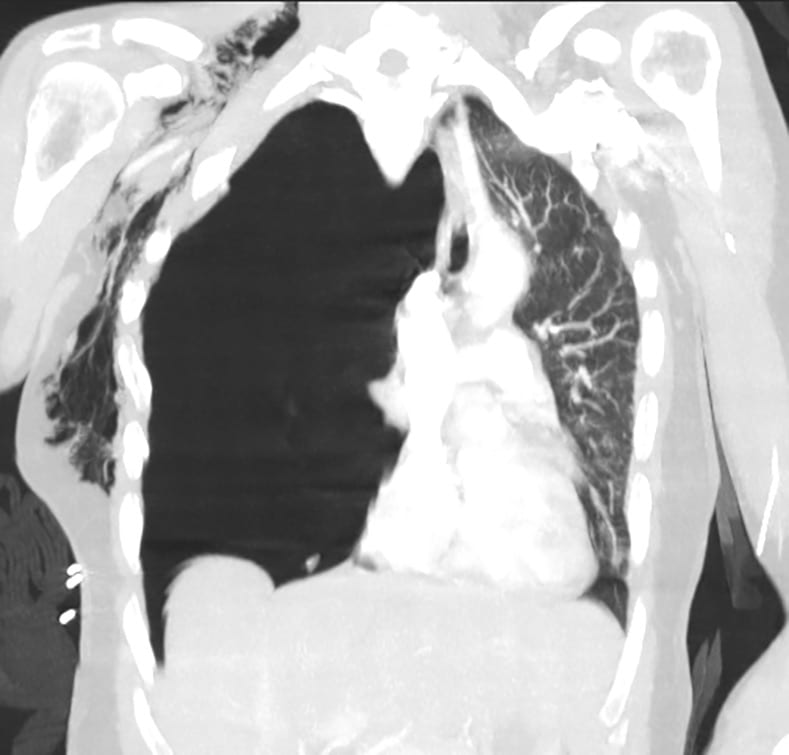
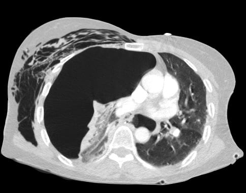
A CT scan is performed. Describe and interpret.
Reveal Answer
The CT scan shows a tension pneumothorax with marked mediastinal shift.
There is associated extensive subcutaneous emphysema. A rib fracture can be seen and is the cause of the pathology.
An intercostal catheter (ICC) is placed and the tension relieved. The first chest x-ray, taken after the intercostal catheter insertion demonstrates resolution of the tension pneumothorax, with the mediastinum returning to the centre of the thorax. Extensive subcutaneous emphysema is evident outlining the pectoral muscles.
24 hours later the patient suffered a further episode of acute dyspnoea with associated mediastinal shift. Follow the rest of the case here…
Related Clinical Cases
- LITFL Ultrasound library
- LITFL TOP 100 ultrasound cases
- LUNG ultrasound WORKED CASES
- LUNG ultrasound LEARNING MODULES

ULTRASOUND LIBRARY
Clinical Cases
An Emergency physician based in Perth, Western Australia. Professionally my passion lies in integrating advanced diagnostic and procedural ultrasound into clinical assessment and management of the undifferentiated patient. Sharing hard fought knowledge with innovative educational techniques to ensure knowledge translation and dissemination is my goal. Family, wild coastlines, native forests, and tinkering in the shed fills the rest of my contented time. | SonoCPD | Ultrasound library | Top 100 | @thesonocave |


