![]()
William Cowper
William Cowper (1666-1709) was an English surgeon and anatomist.
One of the first surgeon-scientists of Great Britain, Cowper was a credible anatomist despite his critics, performing research on significant medical topics and pioneering the application of experimental principles to surgical problems. A man ahead of his time, Cowper’s ideologies would set the foundation for the celebrated Hunterian school of surgery by more than half a century.
Cowper’s works were deemed to be some of the best of his time, having authored two important anatomy books and the first-ever treatise on general physiology for surgeons in the English language. Controversy surrounding plagiarism, however, would stain Cowper’s reputation even to this day. Despite this, he was still one of the first two surgeons to ever be honored by the Royal Society of London.
Cowper is eponymous with Cowper’s Gland and Cowper’s Fluid.
Biography
- Born 1666 in Petersfield, England
- 1682-1691 – Apprentice to barber William Bignall, then John Fletcher
- 1691 – Diploma of the Barber-Surgeons’ Company; license for surgery from the local Anglican Bishop
- 1696 – Elected Fellow of the Royal Society of London (FRS), one of the first two surgeons to ever so alongside Charles Bernard.
- 1702 – Described Cowper’s glands and Cowper’s fluid; proved the existence of capillaries in higher-order mammals.
- 1705 – Identified arteriosclerosis as a pathology, not a normal process of aging; identified the clinical symptoms and basic pathophysiology of aortic stenosis and regurgitation
- Died March 8, 1709 in Bishop Sutton, England
Medical Eponyms
Cowper’s glands and Cowper’s fluid (1699)
Cowper’s glands are also known as the bulbourethral glands. A pair of pea-shaped exocrine glands found posterolateral to the membranous urethra in males which secrete Cowper’s fluid; a fluid rich in mucoproteins that lubricates the distal urethra and neutralizes acidic urine prior to ejaculation.
1684 – Jean Méry (1645-1722) first published a description of the bulbourethral glands
Original
English
M. Mery a découvert dans l’Homme sous la partie virile, deux petites glandes de la grosseur d’un pois. Elles sont, placées au dessous des muscles accélérateurs, et cloignées du corps des Prostates d’environ un poulce. Il y a entre elles une distance d’environ deux lignes.
Mr. Mery discovered two small galnds the size of a pea under the virile part of the man. They are positioned below the accelerator muscles, and separated from the body of the prostate by about a pulse. There is a distance of about two lines between them
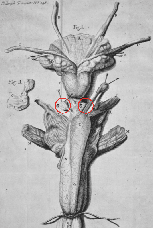
1699 – William Cowper presented the first detailed description of the structure, topography and function of the bulbourethral glands
About a quarter of an Inch below the Prostate Glands, (E) I found two other small Glands (GG) placed on, each side the Urethra (F) a little above the Bulb of its Cavernous Body: These Glands are of a deprest Oval Figure, not exceeding the magnitude of a small French Bean. After those parts of the Musculus Accelerator (L) are removed, which pass over these Glands, you may feel them placed like two hard Bodies on each side the Urethra. Their Excretory Ducts appear on their internal Surface next the inner Membrane of the Urethra whence they descend about half an inch in length before they grow less and pierce that Membrane obliquely at their opening into the Urethra, (D) in which they discharge their separated Liquor….The two glands above described, which from the Liquor they separate may be called ‘Glandula Mucosae’
Cowper 1699
The Artifice of Nature is very extraordinary in thus placing these Glands and their Excretory Ducts, since en the Erection of the Penis and the distension of the Bulb of the Cavernous Body of the Urethra, they are thereby necessarily compressed, and the Liquor contain’d in their Excretory Ducts forced through their two Orifices into the Cavity of the Urethra: besides this, that pare of the Musculus Accelerator which passes over these Glands, contributes to this Compression. It seems requisite such Agents should Conspire in compressing these Organs, since the Liquor they separate is so very Tenacious; which consistence of it is absolutely necessary for the Uses it is employed in.
Cowper 1699
Key Medical Attributions
A man of discoveries
As one of Britain’s first surgeon-scientists, Cowper made a few landmark discoveries.
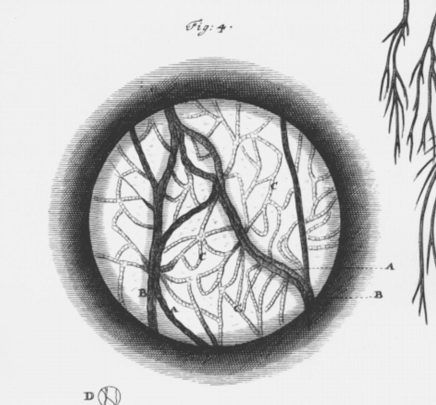
In 1702, Cowper was the first to prove the existence of capillaries in higher-order mammals by using the elegant preparations of mesentery from cats and dogs.
After the circulation of the blood through the heart, lung, and large blood vessels was demonstrated by Dr. Harvey, it was only a guess how the extremities of the arteries transmitted the blood to the veins, till Mr Lewenhueck’s microscopes had discovered the continuation of the extremities of those vessels in fish, frogs, etc. which is now commonly shown by microscopes made by other hands. Yet there are not wanting those who doubt of the like continuations of the extremities of arteries and veins in human bodies and quadrupeds…For this end, I took a young Cat and flattened it to a board…the omentum and intestine were extruded…I saw the globules of the blood move very swiftly in the small vessels, which are only to be seen in the most transparent parts of the membranes of its omentum, but the motion of the blood soon abated, and its globules were withdrawn from the extremities of its blood vessels, and in a little time became stagnant in their larger branches.
Cowper 1702: 1180-1181
In 1705, Cowper reported on the calcific narrowing of arteries. He concluded that they were not normal, but rather a pathological process. He also demonstrated that this was the principal reason that the feet of elderly people became gangrenous. Until then it had been taught that mortifications and gangrenes were caused by the “want of spirits” or the “degeneration of humouis.”
The ossification or petrification in the Great Artery, at it rises from the Heart, has been so commonly found, that some think it is constant; how if may be in some animals I cannot be certain, but in human bodies I am well assured whenever it happens it is a disease, and does in some measure incommode those parts…
Cowper 1705: 1971
The article on calcific narrowing also saw Cowper describe the clinical symptoms and basic pathophysiology of aortic valvular stenosis and aortic regurgitation, making him one of the first to do so.
The symptoms in his Illness plainly showed what must follow, from the disorders of these valves as they are rendered more or less useless: For as their Office is to prevent the return of the blood into the heart…if by any accident they are hindered from doing their duty…the consequences must be, not only a regurgitation of blood into the heart, but they baulk its impulsive force, when the muscular fibers (which are in these valves) cannot contract to prepare the passage for the blood of the left ventricles when to be expelled into the aorta, hence the intermissions of the pulse…may be accounted for…In both these cases the left ventricle of the heart was dilated proportionally to the ill constitution of these valves, which clearly shows these valves give that assistance to the heart in its office that it cannot be without, and that it gradually suffers according to their indisposition.
Cowper 1705: 1973
Cowper’s works over the years were critically acclaimed for their comprehensiveness and lack of systematic theorizing and exaggerated conclusions. His works would set the standard for the surgeon-scientists who came after him.
Major Publications
Also published as Gulielmus Cowper;
- Cowper W. Myotomia reformata, or, A new administration of all the muscles of humane bodies. 1694
- Cowper W. The anatomy of humane bodies. 1698
- Cowper W. An account of two glands and their excretory ducts lately discover’d in human bodies. Philosophical transactions, 1699; 21(258): 364-369 [Cowper’s Glands and Cowper’s Fluid]
- Cowper W. An account of stitching the great tendon, between the calf of the leg and heel, with its union and cure, after an entire division of it. Philosophical transactions, 1699; 21(252): 153-160
- Cowper W. Glandularum quarundam, nuper detectarum, ductuumque earum excretoriorum, descriptio, cum figuris. 1702
- Cowper W. An account of diverse schemes of arteries and veins. 1702; 23(280): 1177-1201
- Cowper W. Of officiations or petrifications in the coats of arteries, particularly in the valves of the great artery. Philosophical transactions, 1705; 24(299): 1970-1977
- Cowper W. Myotomia reformata: or an anatomical treatise on the muscles of the human body. Illustrated with figures after the life. By the late Mr. William Cowper. 1724
Controversies
Cowper as the medical plagiarist
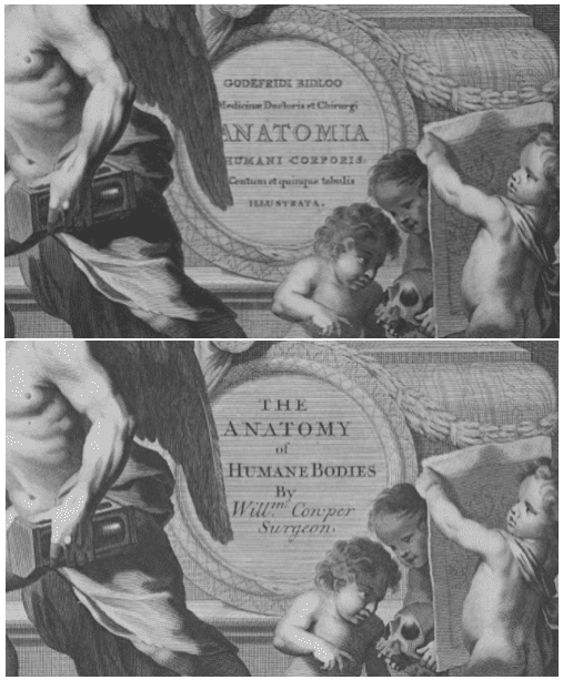
Cowper published the book The Anatomy of Humane Bodies (1698) 13 years after Govard Bidloo (1649 – 1713) published Anatomia Humani Corporis (1685).
Bidloo’s book with 105 plates drawn by Gerard de Lairesse and engraved by Abraham Blooteling did not create much attention or sales, so he sold 300 copies of his engravings to Cowper’s publishers. Cowper was asked to translate Bidloo’s text from Dutch to English, using the same information and engravings.
Cowper added nine new engravings because “…he believed Bidloo’s work failed to properly express or cover relevant information,” and replaced Bidloo’s name and original title with his own. Cowper did not credit Bidloo on the title pages, but placed Bidloo’s name in his introduction.
Clearly Cowper plagiarised Bidloo’s work, as was common at the time. However Cowper’s plagiarism was all the more egregious as he undermined Bidloo’s original work whilst criticising the original author.
In 1700, Bidloo petitioned the Royal Society [Gulielmus Cowper, criminis literarii citatus, coram tribunali] to repeal Cowper’s membership of the Royal Society, however, since Cowper legally purchased the plates from the publisher, there were no retributions for his plagiarism.
References
Biography
- Closterman, J. Portrait: Gulielmus Cowper (1666-1709), Estampe, Gravure de portraits
- Buckmann RF, Futrell JW. William Cowper. Surgery 1986; 99(5); 582-590
- Moore N. Cowper, William (1666-1709). Dictionary of National Biography, 1885-1900, Volume 12
- Sanders MA. William Cowper and his decorated copperplate initials. The Anatomical Record Part B: The New Anatomist 2005; 282B(1): 5–12
- Bibliography. William Cowper (1666 – 1709). World Cat Identities
Eponymous terms
- Mery J. Observations anatomiques. Journal des Sçavans 1684: 201
- Mascari K. William Cowper’s Anatomy of Humane Bodies. SLU Special Collections
Eponym
the person behind the name
Lewis is an RMO at Royal Perth Hospital. He is currently interested in critical care medicine.
BA MA (Oxon) MBChB (Edin) FACEM FFSEM. Emergency physician, Sir Charles Gairdner Hospital. Passion for rugby; medical history; medical education; and asynchronous learning #FOAMed evangelist. Co-founder and CTO of Life in the Fast lane | On Call: Principles and Protocol 4e| Eponyms | Books |