
![]()
William Cumming
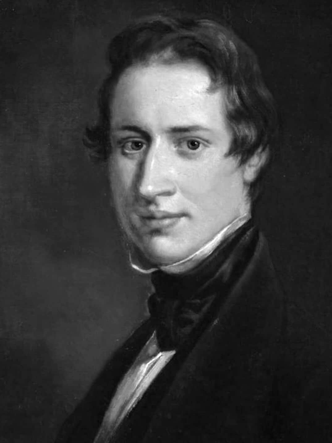
William Cumming (1822-1855) was an English ophthalmic surgeon
Cumming was a gifted ophthalmic surgeon whose work foreshadowed the development of the ophthalmoscope. In 1846, he demonstrated that, under carefully controlled lighting, the human eye’s fundus could reflect light back to an observer, allowing glimpses of the retina and choroid in life. While he never obtained a detailed view of retinal vessels, his insight provided the conceptual foundation upon which Helmholtz later built the first practical ophthalmoscope.
Cumming’s method relied on excluding stray light, placing the patient’s eye at an optimal distance from a light source, and positioning the observer close to the axis of illumination. He recognised the potential for diagnosing retinal and choroidal disease, even recommending pharmacologic dilation with atropine to improve the view decades before mydriasis became standard practice in ophthalmoscopy.
Biographical Timeline
- Born May 9, 1822 in St Anne, Limehouse, Middlesex, England, son of Dr William Spink Cumming
- 1846 – While working at the Royal London Ophthalmic Hospital (Moorfields), presented On a Luminous Appearance of the Human Eye to the Medico-Chirurgical Society of London, describing conditions under which a reflex from the human fundus could be seen.
- 1846–1851 – Discussed his observations with colleagues John Dixon and William Bowman; suggested that the choroid and its pigment were the likely reflecting surface.
- 1851 – Helmholtz publishes Beschreibung eines Augenspiegels, providing a practical method for retinal examination.
- Died June 5, 1855 at Limehouse, aged 33.
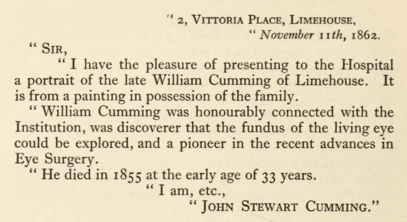
- 1862 – Portrait of Cumming presented to Moorfields Eye Hospital by his family; still displayed in the Board Room.
Key Medical Contributions
Cumming and the Ophthalmoscope
In June 1846, William Cumming presented his landmark paper On a Luminous Appearance of the Human Eye, and its Application to the Detection of Disease of the Retina and Posterior Part of the Eye to the Medico-Chirurgical Society of London. Cumming had noted that a reflex could be obtained from the fundus of the human eye under certain conditions of illumination.
The object of the present paper is to show that the healthy human eye is equally or nearly equally luminous as the eye of the cat, dog, &c., when observed under favourable circumstances, and the application of the abnormal appearance, or want of, this luminosity, to the detection of changes in the retina and posterior part of the eye
Cumming detailed the conditions necessary to observe luminosity:
a. That the eye must be at some distance from the source of light; the distance being greater in proportion to the intensity. b. That the rays of light diffused around the patient (and sometimes around the eye itself) should be excluded. c. That the observer should occupy a position as near as possible to the direct line between the source of light and the eye examined
Cumming then outlined the process by which he was able to observe the retina in the living eye:
The retina and choroid hitherto concealed in the living eye, and little opportunity being afforded of examining their condition after life, in consequence of their diseases not terminating fatally, considerable uncertainty had hitherto attended the diseases ascribed to these structures; but the existence of this luminosity, its non-existence, or abnormal appearance may enable us to detect changes in these structures hitherto unknown, or satisfactorily to see those which we only suspected.
If we dilate the pupil with atropine, we have the means afforded of seeing the condition of the retina and choroid in every case.
Although he lacked the means to visualise the retinal vessels as Helmholtz would later achieve in 1851, Cumming’s work established the foundational principle that light could be reflected from the living human eye and used for diagnosis, an essential conceptual step towards the invention of the ophthalmoscope.
Legacy and Controversies
Ophthalmoscope (1846): Though often miscredited as the ophthalmoscope’s inventor, Cumming’s real achievement was in recognising the diagnostic potential of the fundus reflex. Helmholtz’s 1851 instrument transformed this observation into a reproducible clinical technique.
Which William Cumming? Confusion over his identity has persisted, with some sources conflating him with other contemporaries named William Cumming, including
- William Cumming (1812–1886) – Scottish physician, possibly confused in later references because of overlapping professional scope and time period.
- William Cumming (1769–1852) – Irish painter; entirely unrelated, but frequently mislinked online
- William Spink Cumming (1793-1872) – Scottish physician, father of William Cumming
- William Cumming (1810-1889)
Major Publications
- Cumming W. On a luminous appearance of the human eye, and its application to the detection of disease of the retina and posterior part of the eye. Med Chir Trans. 1846;29:283-96.
References
Biography
- The discovery of the ophthalmoscope In: The history & traditions of the Moorfields Eye Hospital : one hundred years of ophthalmic discovery & development 1929: 100-102 plus portrait plate
- Dixon J. Cumming, William (1822-1855). Dictionary of National Biography, 1885-1900, Volume 13
Eponym
the person behind the name
BA MA (Oxon) MBChB (Edin) FACEM FFSEM. Emergency physician, Sir Charles Gairdner Hospital. Passion for rugby; medical history; medical education; and asynchronous learning #FOAMed evangelist. Co-founder and CTO of Life in the Fast lane | On Call: Principles and Protocol 4e| Eponyms | Books |