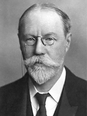
Charles Beevor

Charles Edward Beevor (1854-1908) was an English neurologist
Beevor was a man of exceptional modesty and simplicity of character, self-critical to the highest degree, and gifted musically and artistically
Beevor specialised in the study of neuroanatomy and physiology. He worked closely with neurosurgeon Sir Victor Alexander Haden Horsley (1857-1916) and Charles A. Ballance (1856-1936) on problems of cerebral localisation, devising a novel method to identify cerebral vascular territory maps
His notable publications include his Diseases of the nervous system: a handbook for students and practitioners (1908); his Croonian lectures on muscular movements and their representation in the central nervous system (1903); and the cerebral arterial supply. (1908) which filled a gap in contemporary anatomical knowledge
Remembered for describing the Beevor sign – upward movement of the umbilicus with truncal flexion from a supine position – indicating a spinal cord lesion between T10 and T12 and Beevor’s axiom
Described the jaw-jerk with Armand de Watteville (1846–1925) in 1885
Biography
- Born June 12, 1854 son of prominent English surgeon Charles Beevor, FRCS
- 1878 – MB, University College, London [MRCS 1878, MD, 1881, MRCP 1882; FRCP 1888]
- Following house physician position at the the National Hospital for the Paralysed and Epileptic, he studied at Vienna, Leipzig, Berlin and Paris, his teachers including Carl Weigert (1845-1904), and Wilhelm Heinrich Erb (1840-1921)
- 1883 – assistant physician, rising to physician to the National Hospital for the Paralysed and Epileptic, Queen Square
- 1885-1908 assistant physician, rising to physician at the Great Northern Central Hospital
- 1886 – elected a founding member of the Neurological Society of London [vice president 1905, 1906; president 1907]
- Died suddenly on December 5, 1908 after after attending the annual dinner of the Royal Society of Medicine
Medical Eponyms
Beevor sign (1898)
Upward movement of the umbilicus with truncal flexion from a supine position, associated with a spinal cord lesion between the levels of T10 and T12
1898 – Beevor first described the sign in his in his 1898 textbook Diseases of the Nervous System when discussing the function of the rectus abdominis muscles
The rectus abdominis of either side can be felt to contract by making the patient attempt to get into the sitting position while lying down without using his arms, being careful at the same time to fix the thighs. If the lower halves of the recti alone are paralysed, the umbilicus is drawn upwards in making this movement.
Neurologists praised his sign for its ability to localise spinal cord lesions in the absence of sensory symptoms and its usefulness in distinguishing organic paraplegia from functional or hysterical paraplegia.
Beevor went on to further define the sign in his Croonian Lectures given in 1903 and published in the British Medical Journal and the Lancet
It is often important to know, especially in cases of tumour of the spinal cord, if any part of the recti abdominis are paralysed as they are long muscles which are supplied from the sixth to the twelfth dorsal nerves. I observed some years back a symptom which enables the observer to tell if there is any weakness of the upper or lower parts of the recti. This symptom is the movement of the umbilicus. In health in the movement of sitting up the umbilicus does not alter its position, but if from a lesion of the lower part of the cord or its nerves the part of the recti below the umbilicus is paralysed the normal upper part of the recti draws up the umbilicus sometimes to the extent of an inch. As the abdominal wall at the level of the umbilicus is supplied by the tenth dorsal root any marked elevation of the umbilicus in the act of sitting up would show a lesion between the tenth and twelfth dorsal segments of the cord or the roots coming off from them. In the case where I first observed this symptom there was a malignant growth involving the cord at the level of the eleventh and twelfth dorsal roots.
Since its original description, the Beevor sign has been described in myriad disease processes including spinal cord tumors, facioscapulohumeral muscular dystrophy (FSHD), Lyme disease, acute disseminated encephalomyelitis (ADEM), spinal cord infarction, myotonic dystrophy type 1, and sporadic inclusion body myositis.
Beevor’s Axiom
the brain knows nothing about muscles; it only knows about movements
The axiom, commonly applied in rehabilitation medicine, is attributed to Beevor following his 1898 publication on cerebral localisation
The reason of this is that the movement was required to be to the opposite side; it is immaterial whether the muscle moved was on the same side, or on the opposite side; the brain knows nothing about muscles, only of movements and it is a mechanical convenience that the left sternomastoid should be used to turn the head to the right side.
In 1899, the very eminent John Hughlings Jackson (1835-1911) couched the phrase in similar terms in an address to the British Medical Association
Here I may best remark on the differences between muscles and movements, a matter of vast importance. To speak figuratively, the central nervous system knows nothing of muscles, it only knows movements.
…and from thenceforth, Beevor attributed the axiom to Hughlings Jackson
1902 – It is another illustration of that dictum of Dr. Jackson’s, that the brain knows nothing about muscles, but only about movements. [Beevor 1902]
..if in dealing with hemiplegia we always remember Hughlings Jackson dictum, that the brain knows nothing about muscles but only of movements, and all that we have to take int account is, whether the movement takes place in the field of action on the right or the left of the antero-posterior mid-line of the body. [Beevor 1909]
Jaw jerk (chin reflex, mandibular reflex, masseter reflex)
1882 – Philadelphia neurologist Morris James Lewis (1852-1928) described the ‘chin reflex’. His initial observations were made in 1882 and briefly reported in the Philadelphia Medical News in a paper by Silas Weir Mitchell (1829-1914) On facial neuralgia treated with neurectomy. Subsequently, he
reported further details of the chin reflex in a presentation to the Philadelphia Neurological Society on February 23, 1885
“The Chin Reflex. A New Clinical Observation.” In the winter of 1882, while examining, at the Infirmary for Nervous Diseases connected with the Orthopaedic Hospital, Philadelphia, a case of section of the inferior dental nerve. Dr. Morris J. Lewis discovered a new reflex, [For report of case see Philadelphia Medical News, 1882.] This consists of a sudden elevation of the lower jaw immediately following a blow upon the lower teeth, or chin, and is most easily produced by striking the parts mentioned in a downward direction with a rubber plexor. The mouth of the patient is of necessity open, and the muscles should be relaxed.
1886 – Beevor reported jaw clonus in a woman with “wasted forearm muscles due to amyotrophic lateral sclerosis“. deW appended “the phenomenon was observed in a case in America”
As Dr. Gowers has called my attention to a case published in America, in which an increased tendon-reflex of the muscles of the lower jaw was obtained, I have thought that this case, which I saw four years ago when resident at the National Hospital for the Paralysed and Epileptic, might be of interest.
The symptom which I think the most interesting in this case is the clonus of the lower jaw; this could very readily be produced by placing the finger on the teeth of the lower jaw and then depressing it, when immediately the muscles which closed the jaws passed into a state of clonic contraction, and the lower jaw vibrated as long as the pressure was kept up by the finger on the teeth…Clonus could also readily be produced by striking the masseters…
Armand de Watteville coined the term ‘jaw-jerk’ as an editorial postscript to Beevor’s article
As it does not appear to be generally known that a “jaw-jerk” can be readily elicited by an appropriate stimulus in most healthy persons, I take the opportunity offered by the publication of Dr. Beevor’s observation to call the attention of neurologists to this point. The phenomenon is clearly of the same nature as that of the ” knee-jerk,” and is due to the sudden stretching of the masseter and other muscles of mastication. Hence the name I have ventured to give to it, in preference to the longer and less accurate term mandibular (or masseteric) tendon-reaction (or reflex).
When exaggerated, the jaw-jerk may be elicited by using the fingers to depress and mediately percuss the lower jaw. The left forefinger is placed either upon the teeth…or outside upon the projecting mental ridge, and the right index and middle finger used instead of the hammer
de Watteville, 1886 [note following Beevor publication as editor for the journal Brain]
Major Publications
- Beevor CE. A case of amyotrophic lateral sclerosis with clonus of the lower jaw. (Beevor CE). With a note on the jaw jerk or masseteric tendon reflex reaction in health and disease (De Watterville A.). Brain 1886; 8: 516 –518 [jaw reflex]
- Beevor CE, Horsley V. An experimental investigation into the arrangement of the excitable fibres of the internal capsule of the bonnet monkey (Macacus sinicus) Philos Trans R Soc Lond B 1890; 181B: 49–88.
- Beevor CE. A case of myopathy, facio-scapulo-humeral type. Trans Clin Soc Lond 1895; 28: 245–247
- Beevor CE. Diseases of the nervous system: a handbook for students and practitioners. 1898
- Beevor CE. A lecture on Cerebral Localisation. Clinical Journal. 1898; 17(15): 281-287 [Beevor’s axiom]
- Beevor CE. On cases of myopathy. The Practitioner 1899; 62: 663–670.
- Beevor CE. A lecture on the action of muscles. Clinical Journal. 1902; 20(17): 257–261
- Beevor CE. A demonstration of cases at the Great Northern Central Hospital. Clin J 1902; 20: 189–192.
- Beevor CE. The Croonian Lectures on muscular movements and their representation in the central nervous system. Lancet 1903; 161(4164): 1715-1724
- Beevor CE. The Croonian Lectures on muscular movements and their representation in the central nervous system: delivered before The Royal College of Physicians of London June 1903. 1904
- Beevor CE. On the distribution of the different arteries supplying the human brain. 1908
- Beevor CE. The coordination of single muscular movements in the central nervous system. JAMA 1908; 51: 89–97.
- Beevor CE. The cerebral arterial supply. Brain 1908;30:403–425.
- Beevor CE. Remarks on paralysis of the movements of the trunk in hemiplegia, and the muscles which are affected. Br Med J 1909; 1: 881-884
- Beevor CE. On the distribution of the different arteries supplying the human brain. Philos Trans R Soc Lond B 1909; 200: 1–55.
References
Biography
- Obituary: Charles Edward Beevor, M.D., F.R.C.P.Lond. Br Med J 1908; 2: 1785–1786.
- Tashiro K. Charles Edward Beevor (1854-1908). J Neurol. 2001 Jul;248(7):635-6.
- McCarter SJ, Burkholder DB, Klaas JP, Boes CJ. Charles E. Beevor’s lasting contributions to neurology: More than just a sign. Neurology. 2018 Mar 13;90(11):513-517.
Eponymous terms
- Hughlings Jackson J. On the comparative study of disease of the nervous system. Br Med J 1889; 2: 355-362
- Rijntjes M. Mechanisms of recovery in stroke patients with hemiparesis or aphasia: new insights, old questions and the meaning of therapies. Curr Opin Neurol. 2006 Feb;19(1):76-83. [Beevor’s axiom)
- Compston A. From the archives. The cerebral arterial supply. By Charles E Beevor MD, FRCP. Brain 1908; 30: 403–25. Brain. 2013 Feb;136(Pt 2):362-7.
Beevor sign
- Awerbuch GI, Nigro MA, Wishnow R. Beevor’s sign and facioscapulohumeral dystrophy. Arch Neurol. 1990 Nov;47(11):1208-9.
- Hilton-Jones. Beevor’s sign. Practical Neurology 2004; 4(3): 176-177
- Pearce JM. Beevor’s sign. Eur Neurol. 2005;53(4):208-9.
- Desai JD. Beevor’s sign. Ann Indian Acad Neurol. 2012 Apr;15(2):94-5.
- Althagafi A, Nadi M. Beevor Sign. StatPearls
Jaw reflex
- Weir Mitchell S. On facial neuralgia treated with neurectomy. Medical News (Philadelphia) 1882; XL(10): 257-262 March 11
- Lewis MJ. The chin reflex. A new clinical observation. Journal of nervous and mental disease. 1885; 12: 203-204
- De Watteville A: Note on the jaw-jerk, or masseteric tendon reaction, in health and disease. Brain 1886;8:518-519.
- Lanska DJ. Morris James Lewis (1852-1928) and the description of the jaw jerk. J Child Neurol. 1991 Jul;6(3):235-6.
- Fine EJ, Lohr LA. The chin reflex. Muscle Nerve. 2003 Mar;27(3):386.
- Fine E, Farooq O. The Jaw Jerk Or Masseter Reflex-A History Of Disputed Discovery. Neurology 2021; 96 (15supp).
Eponym
the person behind the name
