


Andrén–von Rosen view
Andrén–von Rosen view is an AP radiographic projection of the paediatric hip obtained with the femora abducted at least 45° and internally rotated. It was described in 1958 by radiologist Lars Andrén and orthopaedic surgeon Sophus von Rosen (1898–1996) to demonstrate the relationship between the proximal femur and acetabulum in suspected congenital hip dislocation.
The principal landmark is the extended long axis of the femoral shaft, later termed the von Rosen line:
- Normal or reduced hip: the axis is directed towards the upper/lateral acetabular margin.
- Dislocated hip: the axis points towards the anterior superior iliac spine (ASIS).
The two femoral-axis lines are interpreted independently and do not need to meet at a defined midline point.
Why was the view developed?
Andrén and von Rosen were dissatisfied with neonatal radiographic methods that relied upon:
- acetabular angles constructed from poorly defined landmarks;
- measurements of the largely cartilaginous proximal femur;
- and numerical distinctions that overlapped widely between normal and abnormal infants.
They wanted a simple, categorical test to determine normal/reduced versus dislocated using landmarks that could be recognised even when the femoral head had not ossified.
How the original view was performed
Andrén and von Rosen advised:
- AP radiograph of the pelvis with femora forcibly abducted to at least 45°;
- internal rotation of the femora and careful positioning during exposure.
How to read the image
| Finding | Extended femoral-shaft axis |
|---|---|
| Normal / reduced | Directed towards the upper or lateral edge of the acetabular wall |
| Dislocated | Directed towards the anterior superior iliac spine |
| Abduction <45° | May falsely resemble dislocation |
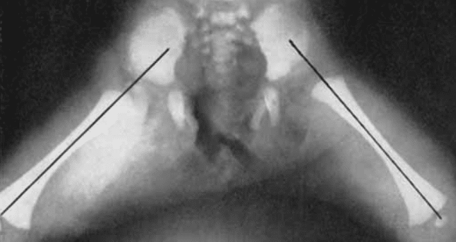
The von Rosen line
The von Rosen line is the extended longitudinal axis of the femoral shaft used to interpret the Andrén–von Rosen view. The term von Rosen line is a later abstraction of the femoral-axis criterion.
It is not a line joining the two femora and it does not require the right and left lines to meet. Each femoral axis is assessed according to its relationship with the ipsilateral acetabulum.
The Andrén–von Rosen view is a positional radiograph in which each extended femoral-shaft axis is judged against the ipsilateral acetabulum. Towards the acetabular margin when reduced, and towards the ASIS when dislocated.
History
1958 – Lars Andrén and Sophus von Rosen published The diagnosis of dislocation of the hip in newborns and the primary results of immediate treatment. They described an AP projection obtained with at least 45° hip abduction and internal femoral rotation. In a normal hip the femoral-shaft axis was directed towards the upper edge of the acetabular wall and in a dislocated hip it pointed towards the anterior superior iliac spine (ASIS).
They demonstrated that insufficient abduction could produce a false impression of dislocation, making accurate positioning fundamental to interpretation.
1962 – von Rosen restated the method in Diagnosis and treatment of congenital dislocation of the hip joint in the new-born. With the femoral head dislocated and the legs internally rotated and abducted approximately 45° the femoral-shaft axis pointed towards the ASIS. After reduction it pointed towards the lateral acetabular margin.
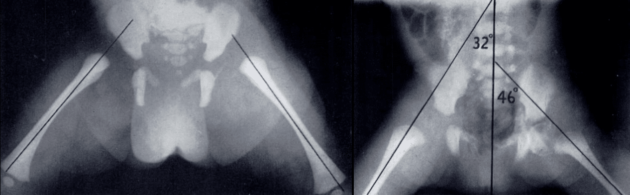
1975 – Knake and Kuhns described a positioning device for the Andrén–von Rosen view after finding that precise bilateral abduction and rotation often required repeat exposures. They concluded that the central long axis of the femur should cross the lateral acetabular margin in an undislocated hip and pass lateral to the acetabulum in a dislocated hip.
1987 – Dietrich Tönnis described the normal extended femoral axis as intersecting the acetabular roof, forming approximately 45° with the sagittal/spinal axis and passing through the body of L4. In subluxation or dislocation, he described the line as passing through the roof edge or above it, forming <40°, and traversing the spine above L3.
These vertebral-level and angular criteria are later refinements, and are not stated in the original 1958 description.
2012 – João Tavares used the abduction–internal rotation (AIR) view to distinguish severe subluxation from true developmental hip dislocation in five children aged 9–20 months. A subluxated femoral head relocated into the acetabulum on the AIR view with restoration or near-restoration of Shenton line but a truly dislocated hip would fail to relocate.
Eponymythology
View first, line later
The von Rosen line was not originally introduced as an independent radiographic line. In 1958 Andrén and von Rosen described the longitudinal femoral-shaft axis as the principal interpretive landmark of their abduction–internal rotation projection.
Later literature increasingly separated the femoral-axis construction from the projection itself, giving rise to the term von Rosen line.
Importantly, the original method assessed the relationship of each femoral axis to its own acetabulum. The two femoral lines were not required to intersect at the midline. Later descriptions by Tönnis added angles to the spinal axis and vertebral levels, but should not be confused with the original 1958 criterion.
Associated Persons
- Erik Sophus von Rosen (1898-1996)
- Dietrich Tönnis (1927-2010)
Alternative names
- von Rosen view
- abduction–internal rotation view
- AIR view
References
Historical references
- Andrén L, Von Rosen S. The diagnosis of dislocation of the hip in newborns and the primary results of immediate treatment. Acta Radiol (Stockh). 1958 Feb;49(2):89-95.
- von Rosen. Diagnosis and treatment of congenital dislocation of the hip joint in the new-born. J Bone Joint Surg Br. 1962 May;44-B:284-91.
- Tönnis D, Legal H, Graf R. Congenital Dysplasia and Dislocation of the Hip in Children and Adults. 1987
Eponymous term review
- Knake JE, Kuhns LR. A device to aid in positioning for the Andrén-von Rosen hip view. Radiology. 1975 Dec;117(3 Pt 1):735-6.
- Tavares JO. Differentiating subluxation from developmental dislocation of the hip. Orthop Rev (Pavia). 2012 Jan 2;4(1):e7.
- Kyle E, Cadogan M. Hip and pelvic lines and arcs. LITFL
eponymictionary
the names behind the name

MB BAO BCh Trinity College Dublin, MSc Clinical Data Analytics University of Galway. Junior doctor in Perth, doctoral researcher in MRI radiomics. Working towards radiology training.

BA MA (Oxon) MBChB (Edin) FACEM FFSEM. Emergency physician, Sir Charles Gairdner Hospital. Passion for rugby, medical history, medical education, and asynchronous learning #FOAMed evangelist. Editor-in-Chief and Webmaster of Life in the Fast lane, LITFL | On Call 4e| Eponyms | Books | Horology |