


Hip and pelvic lines and arcs
Pelvic radiographs contain a network of reference lines, arcs, angles and projected contours. They can be used to assess hip alignment, acetabular development, femoral-head coverage and proximal femoral pathology. Some are simple anatomical constructions whilst others retain the names of the clinicians who described, adapted or popularised them.
No single line should be interpreted in isolation. Their relevance changes with skeletal maturity and the pathology being investigated.
- Developmental dysplasia in the infant and growing hip
- Slipped capital femoral epiphysis (SCFE) during adolescence
- Acetabular coverage and pelvic alignment in the mature hip.
| Clinical setting | Principal lines / signs / measurements |
|---|---|
| Developing Hip | Hilgenreiner, Perkins, Tönnis angle Reimers migration percentage, Andrén–von Rosen view |
| Adolescent / SCFE | Klein line, Trethowan sign, Capener triangle, Shenton line Steel metaphyseal blanch sign, S-sign |
| Mature Hip | Wiberg angle, Sharp angle Köhler line, Köhler teardrop |
Developing / paediatric hip
Assessment of the developing hip depends on reference lines constructed from pelvic landmarks. Hilgenreiner and Perkins provide the basic framework for localisation of the femoral head. Acetabular inclination assesses acetabular development. Reimers quantifies lateral migration and von Rosen provides an additional projectional alignment test.
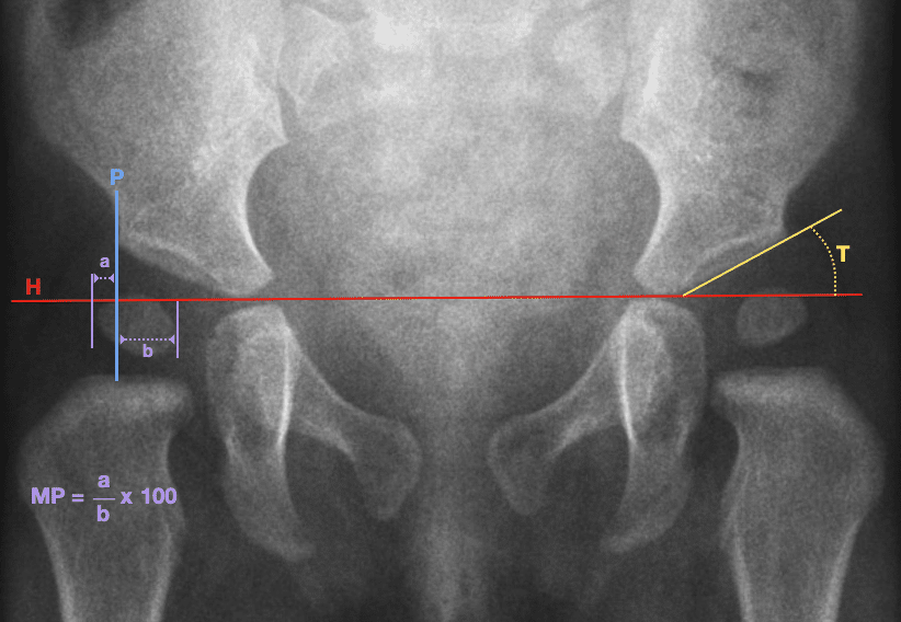
Hilgenreiner line
A horizontal reference line drawn through the triradiate cartilages on the AP paediatric pelvis. It provides the baseline for assessment of developmental dysplasia of the hip and underpins later constructions.
- Heinrich Hilgenreiner (1870–1953), German-Bohemian surgeon
- Hilgenreiner H. Zur Frühdiagnose und Frühbehandlung der angeborenen Hüftgelenkverrenkung. Medizinische Klinik 1925; 21: 1385-1388, 1425-1429
Perkins line
A vertical reference drawn through the lateral acetabular margin, conventionally perpendicular to Hilgenreiner line. Together the two lines divide the developing hip into quadrants and help localise the proximal femur in DDH.
Although now eponymously associated with George Perkins, an equivalent vertical tangent to the lateral acetabular roof had been described by Louis Ombrédanne in 1923, five years before Perkins’ 1928 publication. Later literature also used the terms Ombrédanne line, Putti line and Martin V-line for closely related vertical acetabular references.
- Louis Ombrédanne (1871-1956), French paediatric surgeon
- George Perkins (1892–1979), English orthopaedic surgeon
- Perkins G. Signs by which to diagnose congenital dislocation of the hip. Lancet 1928: 648-650
Tönnis angle
The acetabular roof angle measured between Hilgenreiner line and the acetabular roof. A larger angle indicates a steeper, less developed acetabulum and supports the diagnosis of acetabular dysplasia. Tönnis standardised and popularised the measurement, building on earlier methods of assessing acetabular inclination.
- Dietrich Tönnis (1927-2010)
- Tönnis D, Brunken D. Eine Abgrenzung normaler und pathologischer Hüftpfannendachwinkel zur Diagnose der Hüftdysplasie. Auswertungen von 2294 Pfannendachwinkeln kindlicher Hüftgelenke. Arch Orthop Unfallchir. 1968;64(3):197-228.
Reimers migration percentage
A quantitative measure of how much of the visible femoral head lies lateral to Perkins line on the AP pelvis. Converts hip displacement into a reproducible percentage. Important in hip surveillance, particularly in children with cerebral palsy.
- Jørgen Reimers (1931-2008), Danish paediatric orthopaedic surgeon
- Reimers J. The stability of the hip in children. A radiological study of the results of muscle surgery in cerebral palsy. Acta Orthop Scand Suppl. 1980;184:1-100.
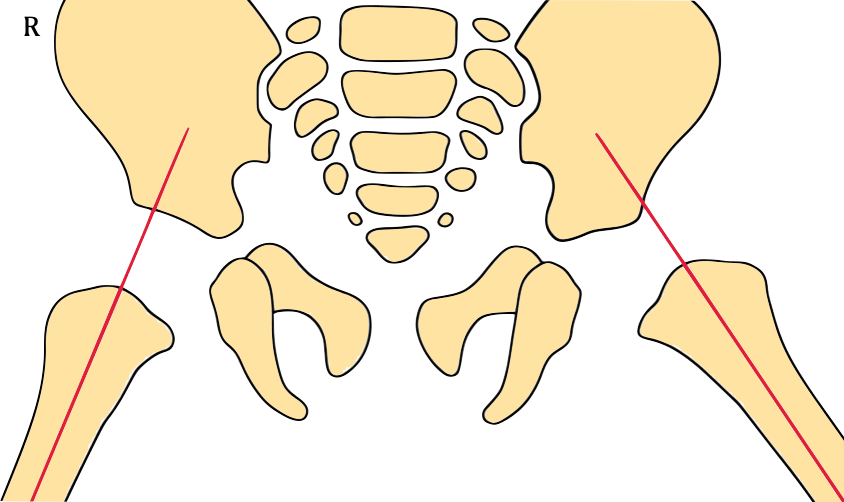
Andrén–von Rosen view / von Rosen line
A specialised AP projection of the neonatal / paediatric hip obtained with the femora abducted at least 45° and internally rotated. The extended femoral shaft axis points towards the acetabular margin in the reduced hip and towards the ASIS in dislocation.
- Erik Sophus von Rosen (1898-1996), Swedish orthopaedic surgeon
- Andrén L, Von Rosen S. The diagnosis of dislocation of the hip in newborns and the primary results of immediate treatment. Acta Radiol (Stockh). 1958 Feb;49(2):89-95.
Shenton line: cross-age alignment sign
A smooth cervico-obturator arc assessing the relationship between the pelvis and proximal femur. Disruption suggests displacement but is non-specific. It can support assessment of DDH, particularly in walking-age children, and remains useful in adolescent and adult hip pathology. Its sensitivity is lower in neonates, where physiological hip positioning makes the arc less reliable.
- Edward Warren Hine Shenton (1872–1955), English radiologist
- Shenton EWH. A diagnostic line about the hip joint. Journal of Physical Therapeutics 1902;3:110-12.
Adolescent hip and slipped capital femoral epiphysis
Radiographic signs of slipped capital femoral epiphysis (SCFE) reflect different consequences of displacement of the capital epiphysis relative to the femoral neck.
On the AP view, Klein line assesses the superior neck–epiphyseal relationship, the Capener triangle reflects acetabular–metaphyseal overlap, and the metaphyseal blanch sign reflects superimposition of the displaced epiphysis and metaphysis. The S-sign assesses contour continuity on the frog-leg lateral projection.
These findings are complementary, and subtle slips may not be apparent from a single AP sign.
Klein – where is the epiphysis relative to the superior neck?
Capener – has normal metaphyseal–acetabular overlap been lost?
Steel – has posterior displacement created metaphyseal superimposition?
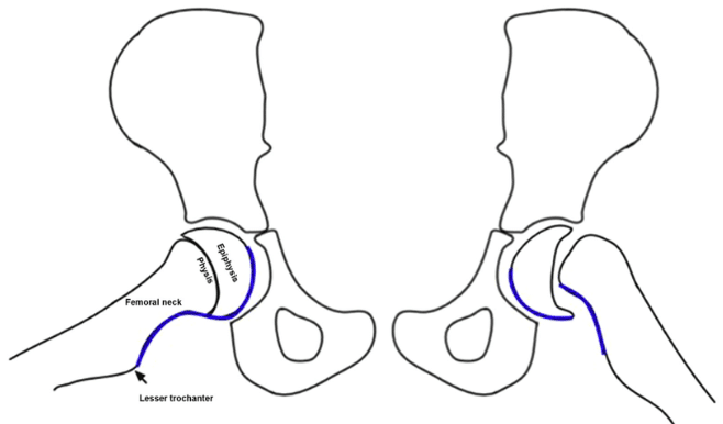
S-sign – has the lateral head-neck contour lost its normal continuity?
Klein line / Trethowan sign
The Klein line is formed by extending the straight superior cortical margin of the femoral neck proximally on the AP radiograph. In the normal growing hip it transects a portion of the lateral capital femoral epiphysis. With early SCFE it may intersect less of the epiphysis than expected, while with greater displacement it may fail to intersect the epiphysis completely.
The Trethowan sign is now commonly used for the abnormal superior femoral neck–epiphyseal relationship demonstrated by Klein line. No primary description by William Henry Trethowan has been located. The earliest attribution identified is W. A. Crabbe in 1968, who described loss of the normal angle between the upper femoral-neck border and lateral edge of the epiphysis.
- Armin Klein (1892–1954), American orthopaedic surgeon
- William Henry Trethowan (1882–1934), English orthopaedic surgeon
- Klein A, Joplin RJ, Reidy JA, Hanelin J. Slipped capital femoral epiphysis; early diagnosis and treatment facilitated by normal roentgenograms. J Bone Joint Surg Am. 1952 Jan;34-A(1):233-9
- Crabbe WA. Trethowan’s sign In: Orthopaedics for the Undergraduate 1968
Capener triangle
The Capener triangle (Capener sign; triangular sign; Scham sign) is the normal dense triangular overlap of the inferomedial femoral neck/metaphysis with the posterior acetabular wall on the AP radiograph. With SCFE, displacement reduces this acetabular–metaphyseal overlap, so the triangle becomes smaller or disappears. It is a supplementary sign rather than a stand-alone diagnostic finding.
Capener did not describe a triangle. He described the developmental anatomy underlying the finding in 1956. Durbin (1960) described the mechanism whilst Jacobs (1962) illustrated and clinically evaluated the triangular overlap. Scham described the “dense triangle” in 1968 and formalised the “triangular sign” in 1974.
- Norman Leslie Capener (1898-1975), English orthopaedic surgeon
- Capener NL. Reconstructive Surgery of the Hip Joint. In: Platt H, ed. Modern Trends in Orthopaedics, 2nd series. 1956: 4-5
- Scham SM. The triangular sign in the early diagnosis of slipped capital femoral epiphysis. Clin Orthop Relat Res. 1974;(103):16-7
Metaphyseal blanch sign / Steel sign
Metaphyseal blanch sign (Steel sign) is a crescent-shaped area of increased density overlying the proximal femoral metaphysis adjacent to the physis on the AP radiograph. It is a projectional overlap produced by posterior displacement of the capital epiphysis behind the femoral-neck metaphysis. The sign may be present before obvious medial displacement makes Klein line abnormal. A lateral radiograph is required when the finding raises suspicion of SCFE.
- Howard Haldeman Steel (1921–2018), American orthopaedic surgeon
- Steel HH. The metaphyseal blanch sign of slipped capital femoral epiphysis. J Bone Joint Surg Am. 1986;68(6):920–922
S-sign
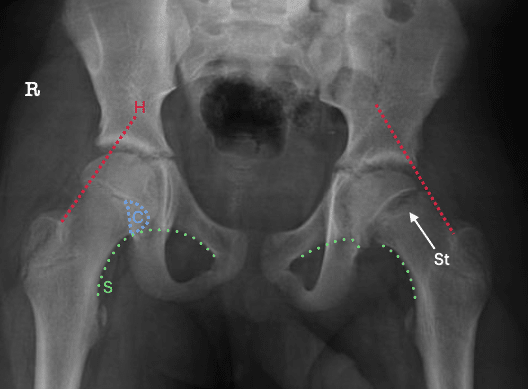
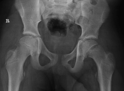
S-sign is a curvilinear sign described on the frog-leg lateral radiograph. The line follows the posteroinferior cortex of the proximal femur from the lesser trochanter, continues along the femoral neck and across the physis, and curves around towards the midpoint of the femoral head. In the normal hip the contour is smooth; broken continuity, asymmetry or a sharp turn is abnormal and may indicate SCFE.
In the original observer study, the S-sign outperformed Klein line and showed better interobserver agreement, although sensitivity remained lower in mild slips.
- Rebich EJ, Lee SS, Schlechter JA. The S Sign: A New Radiographic Tool to Aid in the Diagnosis of Slipped Capital Femoral Epiphysis. J Emerg Med. 2018;54(6):835–843.
Acetabular morphology / mature hip
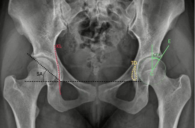
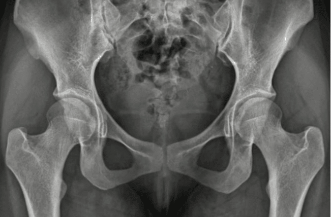
In the skeletally mature hip, pelvic reference lines are useful for assessment of acetabular coverage, orientation, pelvic-column integrity and hip congruity. Quantitative measurements include the centre-edge angle of Wiberg and Sharp angle. Projected anatomical contours include the ilioischial and iliopectineal lines, acetabular walls and teardrop.
Most mature-hip landmarks are anatomical rather than eponymous. The iliopectineal line outlines the anterior pelvic column/pelvic brim, while the anterior and posterior acetabular wall contours help assess acetabular orientation and coverage. Together with the ilioischial line, teardrop and Shenton arc, they form part of the radiographic picture of the AP pelvis.
Centre-edge angle of Wiberg
Centre-edge angle of Wiberg measures lateral acetabular coverage of the femoral head. The angle is formed at the femoral-head centre between a vertical reference and a line to the lateral acetabular roof. Smaller values indicate reduced coverage. Wiberg introduced the measurement in 1939, although both the reference axis and lateral acetabular endpoint have since evolved.
- Gunnar Wiberg (1902–1988), Swedish orthopaedic surgeon
- Wiberg G. Studies on dysplastic acetabula and congenital subluxation of the hip. Acta chirurgica Scandinavica 1939;83(58):5-135.
Sharp angle / Ullmann–Sharp angle
The Sharp angle measures overall acetabular inclination independently of femoral-head position. It is formed between a horizontal line through the inferior pelvic teardrops and a line from the teardrop to the lateral acetabular roof. Increasing values indicate a steeper acetabulum. Sharp described and evaluated the measurement in 1961, although Tönnis later attributed an equivalent construction to Karl Ullmann in 1938.
- Sharp IK. Acetabular dysplasia: the acetabular angle. J Bone Joint Surg Br. 1961;43-B(2):268–272.
Köhler line
Köhler line (ilioischial line) is the projected medial pelvic contour extending from the ilium towards the medial ischium. It provides a reference for medial acetabular depth. The acetabular wall crossing medial to the line indicates a deep acetabulum, while medial projection of the femoral head itself defines radiographic protrusio acetabuli.
- Alban Köhler (1874–1947), German radiologist
- Köhler A. Die normale und pathologische Anatomie des Hüftgelenks und Oberschenkels in röntgenographischer Darstellung. Hamburg: Lucas Gräfe & Sillem; 1905
Köhler teardrop
Köhler teardrop (pelvic teardrop; U-figure) is a U- or teardrop-shaped summation shadow medial to the acetabulum on the AP pelvis. It results from tangential projection of several acetabular and pelvic cortical surfaces. The teardrop is used as a landmark in assessment of acetabular morphology, depth, medial migration and symmetry. Köhler originally called this the Tränenfigur (“tear figure”) and demonstrated that its appearance varied with projection.
- Alban Köhler (1874–1947), German radiologist
- Köhler A. Die normale und pathologische Anatomie des Hüftgelenks und Oberschenkels in röntgenographischer Darstellung. Hamburg: Lucas Gräfe & Sillem; 1905
Eponymythology
The apparent simplicity of pelvic radiographic eponyms conceals a complicated history. Several familiar names were attached retrospectively to observations that their supposed originators did not themselves name. Others describe modifications or reinterpretations of earlier lines, and different traditions have occasionally attached different names to essentially the same radiographic phenomenon.
Perkins and Ombrédanne provide an example of parallel terminology in assessment of the dysplastic hip with the Ombrédanne–Perkins line. Ombrédanne’s 1923 textbook predates Perkins’ 1928 paper and contains the earliest verified description located here of the vertical tangent to the lateral acetabular roof used to assess lateral femoral-head displacement. Perkins subsequently described an independent horizontal–vertical construction which became incorporated with Hilgenreiner line into the familiar modern quadrant system. Later sources collapsed Ombrédanne, Perkins, Putti and Martin terminology into apparent synonymy, obscuring the chronology of the original descriptions.
Klein line and Trethowan sign illustrate later eponymic substitution. The Capener–Scham story provides the clearest example of progressive development. Capener described the underlying anatomy, Durbin described the mechanism, Jacobs illustrated the triangular overlap, and Scham formalised the “triangular sign”.
Conversely, later constructs such as the S-sign show that useful radiographic lines need not acquire an eponym at all.
References
- Campbell SE. Radiography of the hip: lines, signs, and patterns of disease. Semin Roentgenol. 2005 Jul;40(3):290-319.
- Yeap PM, Budak MJ. The pelvic radiograph: lines, arcs and stripes. Singapore Med J. 2021 Jul;62(7):333-340.
- Bi AS, Carter C, Price AE, Litrenta J, Karamitopoulos M, Castañeda PG. A history of eponym usage in hip and pelvis radiography part 1: the paediatric hip. Hip Int. 2023 Mar;33(2):136-143.
- Bi AS, Ryan MK, Castañeda PG, Youm T. A history of eponym usage in hip and pelvis radiography part 2: the adult hip. Hip Int. 2023 Mar;33(2):144-151
eponymictionary
the names behind the name

MB BAO BCh Trinity College Dublin, MSc Clinical Data Analytics University of Galway. Junior doctor in Perth, doctoral researcher in MRI radiomics. Working towards radiology training.

BA MA (Oxon) MBChB (Edin) FACEM FFSEM. Emergency physician, Sir Charles Gairdner Hospital. Passion for rugby, medical history, medical education, and asynchronous learning #FOAMed evangelist. Editor-in-Chief and Webmaster of Life in the Fast lane, LITFL | On Call 4e| Eponyms | Books | Horology |