


Awake in VF
aka ECG Exigency 020
A previously healthy man in his 50s presents to the emergency department with 45 minutes of severe central chest pain. He is alert and orientated. HR 58 bpm, BP 96/60 mmHg, SpO₂ 98% on room air.
This is his ECG:
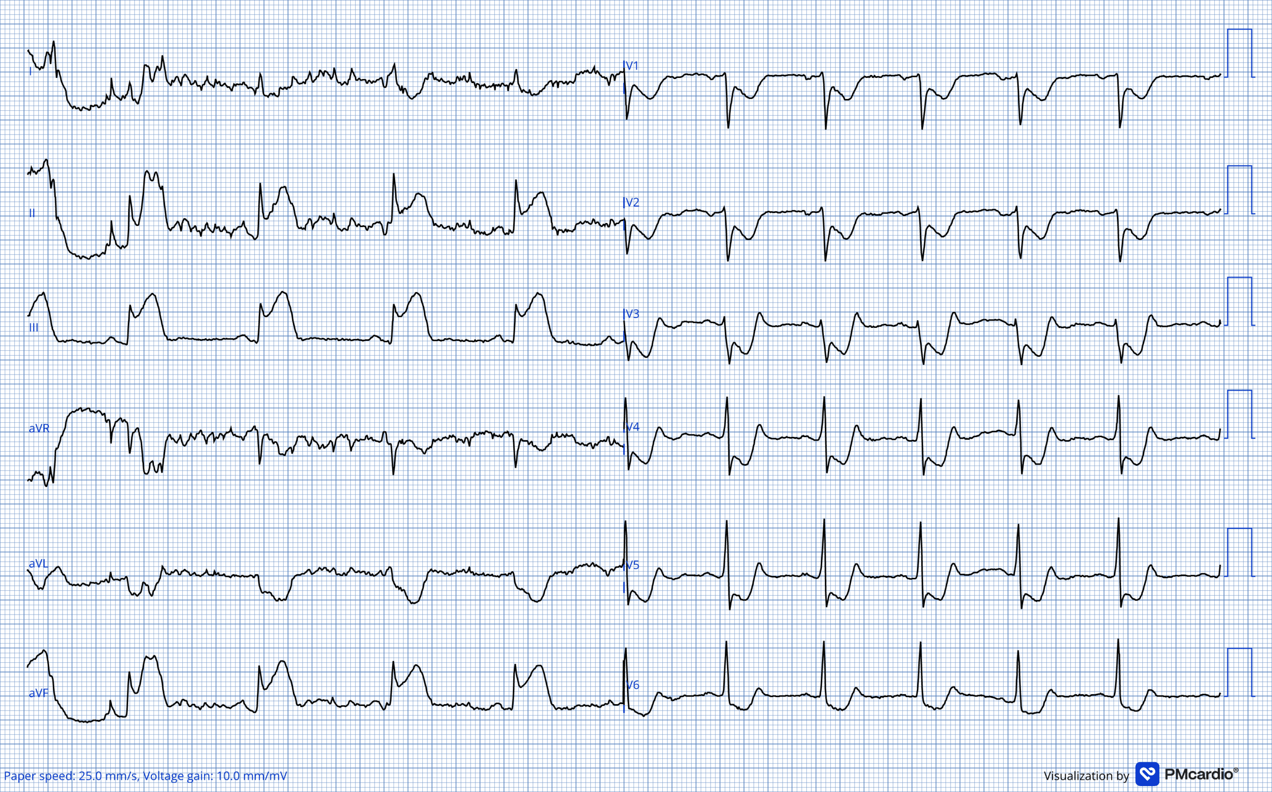
Q1. Describe and interpret the ECG.
Description
- Normal sinus rhythm, rate 60/min
- Marked ST elevation in the inferior leads (II, III and aVF)
- Reciprocal ST depression in I and aVL and anterior ST depression consistent with posterior involvement.
Interpretation
- This is an acute inferior-posterior occlusion myocardial infarction (OMI), most likely due to RCA occlusion.
- The catheter laboratory was activated for immediate reperfusion. Coronary angiography later confirmed an acute thrombotic mid-RCA occlusion.
Note that the 2025 Australian ACS guideline adopts the term acute coronary occlusion myocardial infarction (ACOMI), encompassing both STEMI and STEMI-equivalent ECG patterns. Emphasis has shifted towards pattern recognition rather than relying solely on traditional millimetre-based ST-elevation criteria.
Fifteen minutes later, while preparations for transfer are underway, the patient suddenly collapses into ventricular fibrillation. High-quality CPR is commenced immediately.
Over the next several minutes he remains in VF despite four biphasic defibrillation attempts and intravenous amiodarone 300 mg followed by a further 150 mg. The patient now has refractory ventricular fibrillation.
There is, however, an unexpected complication: during chest compressions he repeatedly opens his eyes and makes purposeful limb movements. No pulse is palpable.
Q2. The patient appears conscious during CPR. Is this ROSC? What should you do?
No. Purposeful movement during chest compressions does not establish ROSC.
CPR-induced consciousness (CPRIC) describes signs of consciousness occurring during cardiac arrest when high-quality chest compressions generate sufficient cerebral perfusion to produce eye opening, speech or purposeful movement. If the rhythm remains VF and there is no evidence of spontaneous circulation, compressions should continue.
Sedation and analgesia may be considered to reduce pain and distress without interrupting CPR. Neuromuscular blockade should not be used simply to abolish signs of CPR-induced consciousness. If paralysis is required for airway management, adequate sedation and analgesia are essential.
In this case, ketamine was administered and the patient was intubated following neuromuscular blockade while CPR continued.
CPR continues without interruption. The rhythm remains ventricular fibrillation.
The initial ECG has already identified the likely cause of the arrest with the presenting ECG strongly supporting acute coronary occlusion. But the patient cannot reach the catheter laboratory while refractory VF continues. Persistent VF now presents two problems: the rhythm itself and the unreperfused coronary occlusion driving it.
At 16:55, during ongoing cardiac arrest, tenecteplase 30 mg IV is administered as a rescue reperfusion strategy.
Q3. What is the role of intra-arrest thrombolysis when acute coronary occlusion is suspected?
Routine thrombolysis is not recommended during cardiac arrest simply because an acute coronary occlusion is suspected.
In PE-related cardiac arrest, systemic fibrinolysis has a recognised guideline role. The evidence is different for coronary thrombosis. Definitive reperfusion is PCI, and randomised trials of empiric intra-arrest thrombolysis such as the TROICA trial have not shown a survival benefit.
In this case, tenecteplase was given as an individualised rescue reperfusion strategy because an acute coronary occlusion had already been demonstrated electrocardiographically and immediate PCI could not be achieved while refractory VF continued. This should be understood as a case-specific decision rather than standard management of VF complicating OMI.
Despite fibrinolysis, ventricular fibrillation persists.
The patient has now received repeated defibrillation, amiodarone, continuous high-quality CPR and treatment directed at the presumed coronary thrombotic substrate. The next problem is persistent electrical instability.
At 16:57, during a rhythm check, an ultrasound-guided left stellate ganglion block is performed using 10 mL of 2% lignocaine.
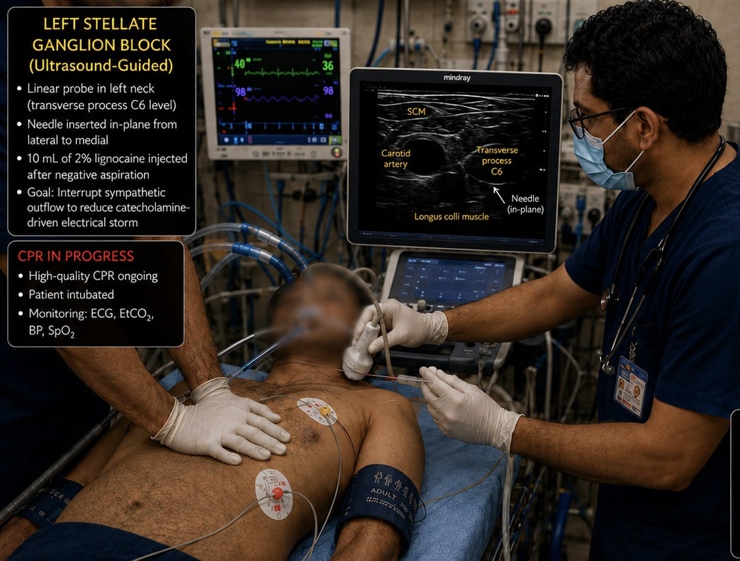
Q4. What is the role of stellate ganglion block in refractory VF management?
Percutaneous stellate ganglion block (SGB) is an emerging rescue therapy for refractory ventricular electrical storm.
Cardiac sympathetic activation can sustain ventricular electrical instability. Left stellate ganglion blockade produces temporary cardiac sympathetic denervation, reducing catecholamine-mediated input and potentially reducing recurrent VT/VF when conventional defibrillation and antiarrhythmic therapy have failed.
Evidence remains predominantly observational. In the multicentre STAR study, 131 patients with refractory electrical storm underwent 184 percutaneous SGB procedures; 92% achieved at least a 50% reduction in treated arrhythmic events during the subsequent 12 hours, with one major complication reported.
SGB should therefore be viewed as an adjunctive rescue strategy, not a replacement for defibrillation, antiarrhythmic therapy or correction of the underlying cause.
Shortly afterwards, an organised rhythm appears and sustained ROSC is achieved at 16:59. Brief AIVR was observed on the monitor following ROSC. Two minutes later, a 12-lead ECG is recorded.
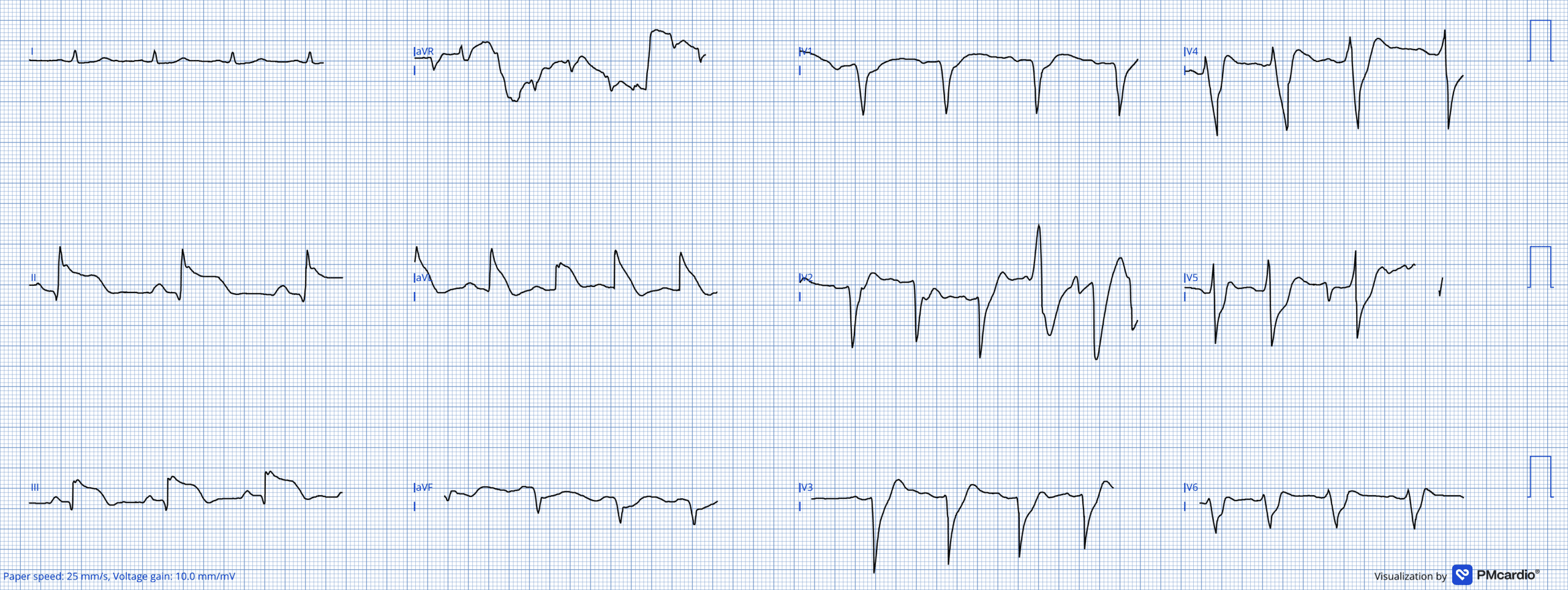
Q5. Describe and interpret the post-ROSC ECG.
The post-ROSC ECG demonstrates an organised rhythm at approximately 70 bpm with persistent changes of acute inferior-posterior myocardial infarction.
There are intermittent broad ventricular complexes with variable morphology and possible fusion/capture beats. These may represent a period of accelerated ventricular activity, but this tracing is not a convincing example of sustained accelerated idioventricular rhythm (AIVR).
More importantly, the ECG does not establish successful coronary reperfusion. AIVR has traditionally been described as a reperfusion arrhythmia, particularly in the thrombolytic era, but it is not a reliable marker of restored epicardial coronary flow. In a contemporary primary-PCI cohort, AIVR was actually associated with less spontaneous ST resolution, less TIMI 3 flow on admission and delayed microvascular reperfusion.
The patient proceeds immediately to coronary angiography.
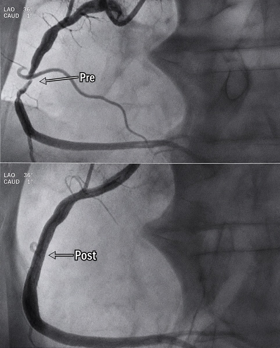
Q6. What does the coronary angiogram show, and how does it relate to the preceding ECGs?
The pre-PCI angiogram demonstrates the acute thrombotic culprit lesion in the mid-right coronary artery (RCA). Following PCI and drug-eluting stent implantation, vessel patency is restored with TIMI 3 flow.
This confirms the RCA culprit predicted by the original inferior-posterior OMI pattern. Importantly, a significant culprit lesion remained and required PCI despite the preceding post-ROSC rhythm, reinforcing that apparent AIVR or other ventricular rhythms should not be taken as evidence of successful epicardial reperfusion.
The patient is extubated the following day, vasopressor support is discontinued and he remains neurologically intact. He is discharged home on day 3 with a modified Rankin Scale score of 0.
The case illustrates the importance of treating both the arrest rhythm and the physiology sustaining it: maintaining perfusion during CPR, addressing the coronary substrate, controlling refractory electrical instability and proceeding to definitive reperfusion.
Learning points
- Inferior OMI can deteriorate rapidly into primary VF before reperfusion is achieved.
- CPR-induced consciousness does not equal ROSC. Apparent consciousness should not interrupt effective chest compressions.
- Persistent VF should trigger continued treatment of the underlying cause as well as the rhythm.
- Intra-arrest thrombolysis for suspected coronary occlusion is not routine therapy and should be distinguished from fibrinolysis for suspected PE.
- Stellate ganglion block is an emerging rescue adjunct for refractory ventricular electrical storm.
- AIVR may occur following reperfusion therapy, but should not be used as proof of restored epicardial coronary flow.
Acknowledgements
Case development, formatting and referencing
- Dr Vinayak M. S., Assistant Professor, Department of Emergency Medicine, Government Medical College, Thiruvananthapuram
- Dr Kathyayini V. R., Assistant Professor, Department of Emergency Medicine, Government Medical College, Thiruvananthapuram
- Dr Roopasree Sivam, Assistant Professor, Department of Emergency Medicine, Government Medical College, Thiruvananthapuram
- Dr Jiss V. Peter, Assistant Professor, Department of Emergency Medicine, Government Medical College, Thiruvananthapuram
Resuscitation team
- Dr Sonu, Junior Resident, Department of Emergency Medicine, Government Medical College, Thiruvananthapuram
- Dr Anagha V., Junior Resident, Department of Emergency Medicine, Government Medical College, Thiruvananthapuram
Coronary angiography
- Dr Cibu Mathew, Professor, Department of Cardiology, Government Medical College, Thiruvananthapuram
Additional contributors
- Dr Jayakumari C., Professor, Department of Emergency Medicine, Government Medical College, Thiruvananthapuram
- Dr Deepa Franklin, Professor, Department of Emergency Medicine, Government Medical College, Thiruvananthapuram
- Dr Anju Mariyam Jacob, Professor, Department of Emergency Medicine, Government Medical College, Thiruvananthapuram
- Dr Chithra V. R., Professor, Department of Emergency Medicine, Government Medical College, Thiruvananthapuram
References
- Australian and New Zealand Committee on Resuscitation. Guideline 11.2 – Protocols for Adult Advanced Life Support. Accessed 7 August 2026. Guideline
- Australian and New Zealand Committee on Resuscitation. Guideline 11.4 – Electrical Therapy for Adult Advanced Life Support. Accessed 7 August 2026. Guideline
- International Liaison Committee on Resuscitation. Consciousness During CPR: ALS Task Force Scoping Review. 2021. ILCOR review
- Olaussen A, Nehme Z, Shepherd M, Jennings PA, Bernard S, Mitra B, Smith K. Consciousness induced during cardiopulmonary resuscitation: An observational study. Resuscitation. 2017 Apr;113:44-50.
- Howard J, Lipscombe C, Beovich B, Shepherd M, Grusd E, Nudell NG, Rice D, Olaussen A. Pre-hospital guidelines for CPR-Induced Consciousness (CPRIC): A scoping review. Resusc Plus. 2022 Nov 28;12:100335
- Böttiger BW, Arntz HR, Chamberlain DA, Bluhmki E, Belmans A, Danays T, Carli PA, Adgey JA, Bode C, Wenzel V; TROICA Trial Investigators; European Resuscitation Council Study Group. Thrombolysis during resuscitation for out-of-hospital cardiac arrest. N Engl J Med. 2008 Dec 18;359(25):2651-62.
- Savastano S et al. Electrical storm treatment by percutaneous stellate ganglion block: the STAR study. Eur Heart J. 2024 Mar 7;45(10):823-833.
- Burns E, Buttner R. Accelerated Idioventricular Rhythm (AIVR). LITFL ECG Library.
- Terkelsen CJ, Sørensen JT, Kaltoft AK, Nielsen SS, Thuesen L, Bøtker HE, Lassen JF. Prevalence and significance of accelerated idioventricular rhythm in patients with ST-elevation myocardial infarction treated with primary percutaneous coronary intervention. Am J Cardiol. 2009 Dec 15;104(12):1641-6.
CLINICAL CASES
ECG Exigency

MBBS (Calicut) MD (AIIMS New Delhi) DNB (Emergency Medicine) MNAMS MRCEM (UK) AHA ACLS Instructor. Emergency physician and Assistant Professor, Government Medical College Thiruvananthapuram. Passion for resuscitation, POCUS, toxicology, critical care, medical education, and simulation. FOAMed enthusiast and creator of EM Rounds with PK sharing practical emergency medicine, evidence, and bedside learning.

MBBS FACEM DDU (Emergency) CCPU. Emergency Physician in Melbourne, Australia. Co-Ultrasound Lead for Emergency Medicine at The Alfred Hospital. Special interests in diagnostic and procedural ultrasound, medical education, and ECG interpretation | ECG Library |
great case, enlightening! thanks you very much! PS would you considered double sequential defib in cases of refractory VF?
Thank you for your comment! Yes, we have used double sequential defibrillation quite often for refractory VF, but unfortunately, the results in our experience were not very reassuring.
Just curious …If SGB had been performed before thrombolysis, the interesting question would be whether it could have broken the electrical storm sufficiently to buy time for reperfusion?
That’s a great question.
The patient was in cardiac arrest with refractory ventricular fibrillation/ventricular tachycardia, and our decision was based on the clinical situation rather than on an established guideline recommending this combination.
Thrombolysis in cardiac arrest remains an area of uncertainty. Trials such as TROICA have not demonstrated a clear benefit for routine fibrinolysis in undifferentiated cardiac arrest. However, in our patient, the arrest occurred in the setting of a strongly suspected acute STEMI, and therefore we considered coronary occlusion as a potentially reversible cause.
At the same time, the patient had refractory VF/VT despite multiple defibrillation attempts and antiarrhythmic therapy. Stellate ganglion block was therefore considered as a rescue therapy for electrical storm. Importantly, there is currently no universally accepted guideline specifying exactly when SGB should be performed during ongoing cardiac arrest. In practice, it is generally considered after recurrent/refractory ventricular arrhythmias despite defibrillation and antiarrhythmic therapy.
The question is therefore whether we should have performed SGB first, waited for ROSC, and then proceeded to primary PCI. That would certainly be a reasonable strategy. However, in this particular situation, we were uncertain whether SGB alone would achieve ROSC, and there was a concern that prolonged refractory VF could deteriorate into asystole or otherwise become non-survivable.
Therefore, our approach was to simultaneously address two potentially reversible mechanisms—coronary occlusion and sympathetic-mediated electrical instability—rather than waiting for ROSC before addressing the coronary lesion.
We acknowledge that this approach is not supported by a specific guideline or high-level evidence, and we do not propose it as a standard strategy. Rather, it represents a physiology-guided rescue approach in an exceptionally challenging clinical scenario. More evidence is needed to determine the optimal timing and sequencing of SGB, fibrinolysis, and coronary reperfusion in refractory ventricular arrhythmias associated with acute STEMI.
Thank you, Dr Roshan, for the detailed clarification. I think your explanation actually highlights the key physiological dilemma very well.
What I find particularly interesting is the possibility that the two interventions may be complementary rather than sequential—SGB addressing the autonomic component of the electrical storm while reperfusion addresses the underlying reversible cause..
It would be interesting to know whether, in your experience, there are any clinical or ECG features that might help identify pattern that can respond to SGB?
Also I appreciate your point that this remains physiology-guided rescue therapy rather than an evidence-based standard, but it certainly raises an important hypothesis for further study..
N.B : And, above all, immense respect for the extraordinary resuscitation effort in such an exceptionally challenging situation—the persistence, clinical judgment, and willingness to pursue every possible reversible cause are truly commendable.
Thank you so much for the recognition and appreciation. Yes, I have performed around 15 stellate ganglion blocks so far. One interesting observation from my experience is that patients with refractory VT or ventricular arrhythmias secondary to acute coronary syndrome (ACS) appear to have a better response to stellate ganglion block compared with patients presenting with ventricular storm in the setting of established structural heart disease.
The underlying physiology may also explain this difference. In ACS-related ventricular arrhythmias, heightened sympathetic activation appears to play an important role in triggering and perpetuating the electrical storm. Stellate ganglion block attenuates this sympathetic drive and may therefore have a particularly useful role in this setting. In contrast, ventricular arrhythmias associated with established structural heart disease may involve a more complex and heterogeneous substrate, including scar-mediated re-entry and persistent electrophysiological abnormalities, where sympathetic modulation alone may be insufficient.
Of course, this is an observation from my clinical experience and would need to be evaluated systematically in larger studies. But it is an interesting physiological distinction and potentially an important area for further research.