


Baumann angle
Baumann angle is an anteroposterior radiographic measurement of coronal alignment of the paediatric distal humerus. It is used principally after reduction of a supracondylar humeral fracture to identify residual varus or valgus malalignment and estimate the risk of subsequent cubitus varus.
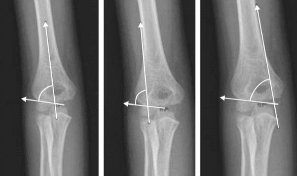
Baumann originally measured the angle between the longitudinal axis of the humerus and a line following the straight lateral distal metaphyseal or capitellar physeal border. A normal original Baumann angle is approximately 64–81°, with a mean near 72°; an increased angle indicates varus malalignment.
A complementary angle between the capitellar physis and a line perpendicular to the humeral shaft is also frequently called the Baumann angle. It equals 90°−α, usually measures approximately 9–26°, and decreases with varus. The measurement convention should therefore always be stated.
Modern use and context
The Baumann angle assesses the coronal-plane orientation of the distal humeral fragment. It is most useful on immediate post-reduction and follow-up radiographs of paediatric supracondylar fractures, when pain, swelling and restricted extension prevent reliable clinical measurement of the carrying angle. It is an alignment and reduction measure rather than a method for diagnosing the fracture itself.
The original shaft–physeal angle is measured on an adequate AP radiograph:
- Draw a line along the longitudinal axis of the humeral shaft.
- Draw a line along the straight lateral portion of the capitellar physis or distal metaphyseal border.
- Measure the proximal angle between the two lines.
| Measurement | Lines used | Reference value | Varus malalignment |
|---|---|---|---|
| Original Baumann angle | Humeral shaft axis and lateral physeal line | 64–81° (95% range) Mean 72° | Angle increases |
| Complementary Baumann angle | Perpendicular to shaft axis and lateral physeal line | 9–26° | Angle decreases |
Because normal alignment varies between individuals, the injured elbow may be compared with the contralateral side when reduction is uncertain. A side-to-side difference of approximately 5° may be significant, but this lies close to the recognised measurement variability of about 5–7°.
Important limitations
The Baumann angle is related to, but does not equal, the clinical carrying angle. Baumann believed that 90°−α represented the carrying angle, but subsequent studies demonstrated a more complex relationship. The post-reduction angle nevertheless correlates with the eventual carrying angle and remains useful for identifying coronal malalignment.
Radiographic positioning is critical. Camp and colleagues found that the measured angle changed by approximately 6° for every 10° of humeral rotation. It should therefore be interpreted alongside image quality, medial-column alignment and the wider clinical and radiographic assessment.
History
1929 — Ernst Baumann (1890–1978) published his original description in Beiträge zur Kenntnis der Frakturen am Ellbogengelenk. Baumann defined angle α between the humeral axis and a straight line passing through the distal lateral metaphyseal border opposite the developing capitellar and lateral trochlear ossification centre.
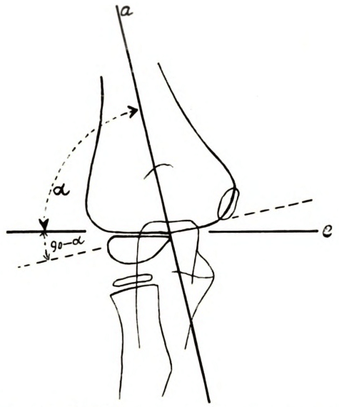
He used the measurement to quantify coronal angulation after supracondylar fracture and to estimate the magnitude of existing or potential cubitus varus. Baumann considered approximately 75–80° normal, allowing for minor individual variation, and suggested that the opposite elbow could be radiographed when greater precision was required.
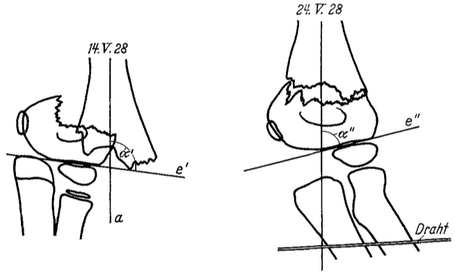
Baumann used serial measurements to assess reduction. In his Figure 17, an angle greater than 90° before treatment returned towards the value of the unaffected elbow following traction.
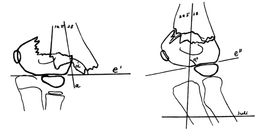
Baumann regarded post-traumatic cubitus varus principally as a malunion deformity rather than a secondary growth disturbance. In 1960, drawing on follow-up of more than 100 cases, he stated that the position of the healed fragments remained unchanged after union and argued that “secondary cubitus varus never develops; it is always the consequence of incomplete reduction.”
1960 – Baumann reaffirmed his 1929 radiographic method in Zur Behandlung der Knochenbrüche am Ellenbogengelenk. He rejected the contemporary suggestion that the short distal fragment could not be assessed accurately. Baumann stated that a line through the lateral condylar epiphyseal zone permitted reliable evaluation of angular displacement, reduction and subsequent development.
This auxiliary line should form a 75–80° superolateral angle with the humeral shaft and the contralateral elbow could be used for precise comparison. Drawing on follow-up of more than 100 cases, Baumann also maintained that post-traumatic “cubitus varus never develops; it is always the consequence of incomplete reduction.”
Baumann restated the measurement he had first described in 1929:
Original
English
Eine Gerade durch die Epiphysenzone des Condylus lateralis gestattet eine zuverlässige Beurteilung der Dislocatio ad axin, der Reposition und der späteren Entwicklung. Diese Hilfsgerade soll oben außen einen Winkel von 75–80° mit der Achse des Humerus bilden.
A straight line through the epiphyseal zone of the lateral condyle permits reliable assessment of angular displacement, reduction and subsequent development. This auxiliary line should form a superolateral angle of 75–80° with the axis of the humerus.
1972 — Hugh S. Dodge incorporated routine measurement of the angle during treatment of displaced paediatric supracondylar fractures with Dunlop traction. Dodge showed that radiographic assessment of coronal alignment after reduction could identify fractures at risk of cubitus varus. This work helped establish the Baumann angle as a practical treatment measure rather than a simple geometrical observation.
1986 — Peter Worlock studied the angle in normal elbows and following reduction of supracondylar fractures. In his publication on the assessment of cubitus varus by the Baumann angle he found a significant relationship between the Baumann angle and carrying angle in normal arms. The angle measured after fracture reduction also correlated with the final carrying angle at follow-up, although it was not simply equal to 90° – α, as Baumann had originally proposed.
1992 — Williamson et al measured the original shaft–physeal angle in 114 normal children aged 2–13 years to assess the Normal characteristics of the Baumann (humerocapitellar) angle. They found mean 72°, standard deviation 4°; and 95% range 64–81°. No significant variation was found with age or sex within the studied population.
1993 — Camp et al examined a paediatric cadaver elbow at progressive increments of internal and external rotation to determine Alteration of Baumann’s angle by humeral position. They demonstrated that humeral rotation altered the measured Baumann angle by approximately 6° for every 10° of rotation emphasising the need for a reproducible AP projection.
2001 — Acton and McNally published “Baumann’s confusing legacy” and compared Baumann’s original publications with later orthopaedic textbooks. They found three different measurements bearing his name:
- the original angle between the humeral axis and lateral physeal line;
- its complement, measured from a perpendicular to the humeral axis; and
- a distal-humeral angle using reference points around the trochlea and lateral physis.
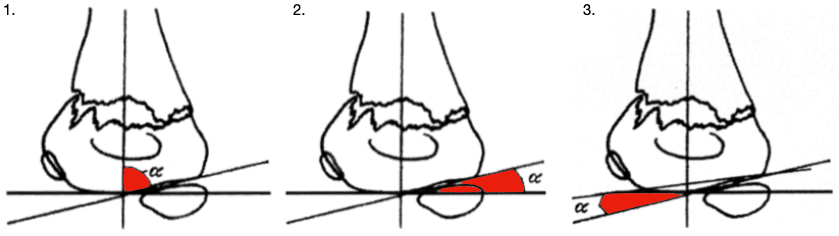
A: Baumann’s original shaft–physeal angle.
B: The complementary angle measured from a line perpendicular to the humeral shaft.
C: A distal-humeral-only angle that does not assess alignment of the distal fragment relative to the shaft. Adapted from Acton and McNally, 2001.
Acton and McNally concluded that Baumann’s original shaft–physeal angle was the historically correct construction. The complementary angle remained geometrically valid if used consistently, but the distal-humeral-only measurement did not assess alignment of the distal fragment relative to the shaft and should not bear Baumann’s name.
2010 — Silva and colleagues published Inter- and intra-observer reliability of the Baumann angle of the humerus in children with supracondylar humeral fractures. They evaluated five observers measuring 35 paediatric elbow radiographs. They used the complementary form of the angle—the lateral physeal line measured against a perpendicular to the humeral shaft.
Interobserver and intraobserver correlations were 0.78 and 0.80 respectively. Repeated or independent measurements commonly differed by as much as 7°, demonstrating that small apparent changes may reflect measurement variability rather than true displacement.
2012 — Krengel and colleagues posed the question Does using the medial or lateral humeral line improve reliability of Baumann angle measurement on plain X-ray? They compared the estimated central humeral axis with lines drawn along the medial and lateral humeral cortices.
Medial and lateral cortical lines improved intraobserver consistency, but the conventional central-axis method retained the best interobserver reliability. Reliability improved when at least 7 cm of the distal humerus was visible on the radiograph.
Eponymythology
Defying Stigler’s law of eponymy, the reviewed literature supports Ernst Baumann as the originator of the measurement attached to his name. The historical controversy concerns definition rather than priority: subsequent authors applied “Baumann angle” to Baumann’s original shaft–physeal angle, its geometrical complement and, occasionally, a third distal-humeral construction.
The most precise terminology is original Baumann shaft–physeal angle for the angle between the humeral shaft and lateral physeal line. The complementary measurement remains clinically useful when clearly identified, but the convention and expected direction of abnormality must always be stated.
Associated Persons
- Ernst Baumann (1890-1978)
Alternative names
- Baumann’s angle
- Baumann humerocapitellar angle
- Shaft–physeal angle
- Complementary Baumann angle — when specifically measuring from the perpendicular to the humeral shaft
References
Historical references
- Baumann E. Beiträge zur Kenntnis der Frakturen am Ellbogengelenk. Unter besonderer Berücksichtigung der Spätfolgen. I. Allgemeines und Fractura supra condylica. Bruns’ Beiträge zur klinischen Chirurgie 1929; 146: 1-50
- Baumann E. Zur Behandlung der Knochenbrüche am Ellenbogengelenk [On the treatment of fractures of the elbow joint]. Langenbecks Archiv für Klinische Chirurgie vereinigt mit Deutsche Zeitschrift für Chirurgie. 1960; 295: 300-4.
Eponymous term review
- Dodge HS. Displaced supracondylar fractures of the humerus in children–treatment by Dunlop’s traction. J Bone Joint Surg Am. 1972 Oct;54(7):1408-18.
- Worlock P. Supracondylar fractures of the humerus. Assessment of cubitus varus by the Baumann angle. J Bone Joint Surg Br. 1986 Nov;68(5):755-7.
- Williamson DM, Coates CJ, Miller RK, Cole WG. Normal characteristics of the Baumann (humerocapitellar) angle: an aid in assessment of supracondylar fractures. J Pediatr Orthop. 1992 Sep-Oct;12(5):636-9
- Camp J, Ishizue K, Gomez M, Gelberman R, Akeson W. Alteration of Baumann’s angle by humeral position: implications for treatment of supracondylar humerus fractures. J Pediatr Orthop. 1993 Jul-Aug;13(4):521-5
- Biyani A, Gupta SP, Sharma JC. Determination of medial epicondylar epiphyseal angle for supracondylar humeral fractures in children. J Pediatr Orthop. 1993 Jan-Feb;13(1):94-7.
- Acton JD, McNally MA. Baumann’s confusing legacy. Injury. 2001 Jan;32(1):41-3.
- Silva M, Pandarinath R, Farng E, Park S, Caneda C, Fong YJ, Penman A. Inter- and intra-observer reliability of the Baumann angle of the humerus in children with supracondylar humeral fractures. Int Orthop. 2010 Apr;34(4):553-7.
- Krengel WF 3rd, Wiater BP, Pace JL, Jinguji TM, Bompadre V, Stults JK, Schmale GA. Does using the medial or lateral humeral line improve reliability of Baumann angle measurement on plain x-ray? The effect of humeral length visualized on the x-ray. J Pediatr Orthop. 2012 Jun;32(4):373-7.
eponymictionary
the names behind the name

MBChB - Liverpool. RMO in Emergency Medicine at Sir Charles Gairdner Hospital, currently soaking up the sun down under. Aiming to pursue a career in anaesthetics.

BA MA (Oxon) MBChB (Edin) FACEM FFSEM. Emergency physician, Sir Charles Gairdner Hospital. Passion for rugby, medical history, medical education, and asynchronous learning #FOAMed evangelist. Editor-in-Chief and Webmaster of Life in the Fast lane, LITFL | On Call 4e| Eponyms | Books | Horology |