


Blunt cerebrovascular injury
BCVI: clinical approach
Blunt cerebrovascular injury (BCVI) is frequently clinically silent until cerebral ischaemia develops. Screen patients with clinical evidence of vascular injury or an appropriate high-risk injury pattern using CT angiography.
Classify confirmed injuries using the Biffl scale and commence antithrombotic therapy as soon as the competing risk of traumatic haemorrhage permits. Repeat vascular imaging according to local protocol, with early involvement of trauma, stroke and neurointerventional specialists for symptomatic, progressive or high-grade injuries.
The following local pathway is based on the Expanded Denver screening criteria and adapted to the imaging resources available at Kelowna General Hospital. Special thanks to Drs M. Moore (Stroke Neurologist), S. Watson (ED and stroke fellow) and S. Nugent (ED and Trauma)
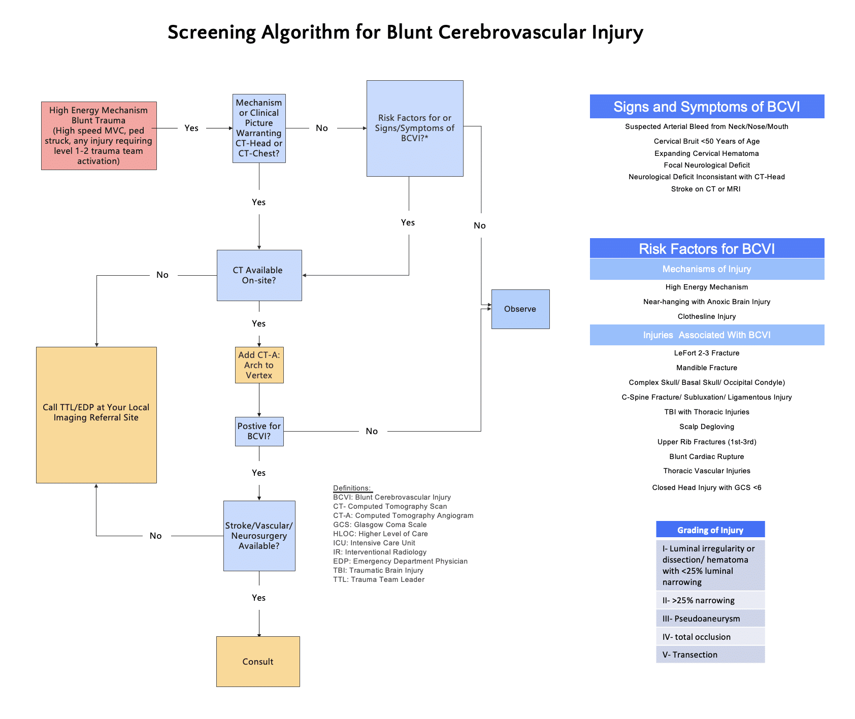
Overview
Blunt cerebrovascular injury encompasses non-penetrating traumatic injury to the carotid or vertebral arteries. Lesions include intimal injury or dissection, intramural haematoma, pseudoaneurysm, occlusion and complete transection. BCVI is identified in approximately 1–3% of patients with blunt trauma, with substantially higher rates among selected high-risk populations.
For most patients, the principal danger is not immediate haemorrhage but delayed cerebral ischaemia caused by thrombosis, distal embolisation or progressive vessel occlusion. Stroke commonly occurs during the first 72 hours after injury and may precede focal neurological or overt vascular signs. Structured screening increases detection, while early antithrombotic treatment is associated with substantially lower rates of stroke and mortality than no treatment.
Mechanism, anatomy and clinical clues
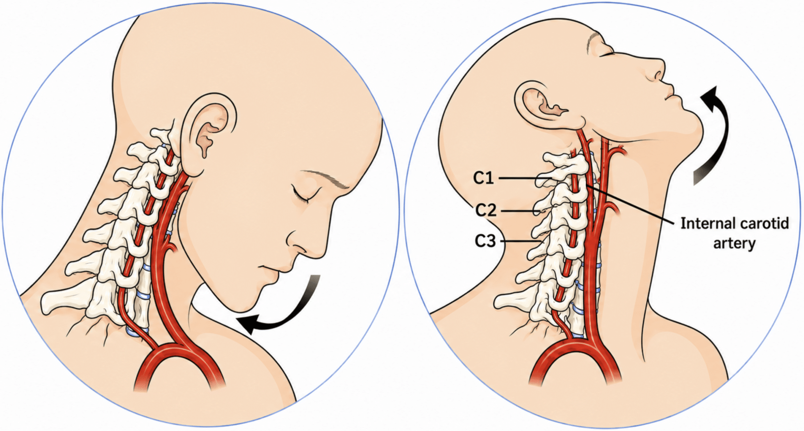
Blunt cerebrovascular injury results from stretching, compression or direct disruption of the carotid or vertebral arterial wall. Hyperextension and rotation may tether the distal internal carotid artery against the upper cervical vertebrae, while flexion, distraction and rotational forces may injure the vertebral artery as it traverses the transverse foramina. Direct neck trauma, intraoral injury and fractures involving the skull base or cervical spine provide additional mechanisms.
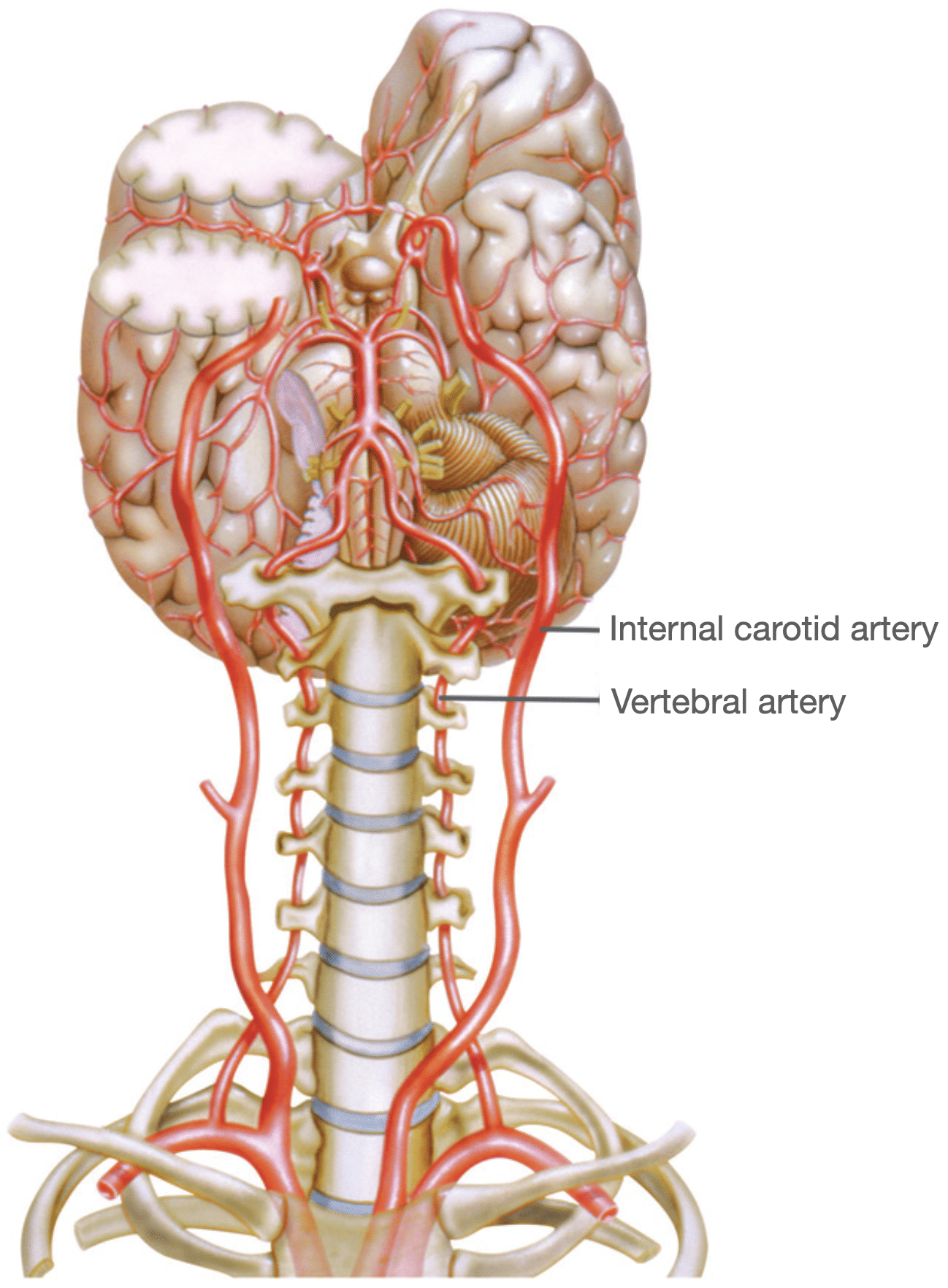
The extracranial internal carotid artery is particularly vulnerable where it is relatively mobile and less well protected in the upper neck. The vertebral artery is at greatest risk where it passes through the cervical transverse foramina and around the upper cervical spine. Cervical spine fractures, subluxation and ligamentous injury are therefore important markers of vertebral artery injury.
Clinical presentation
Most BCVIs are clinically occult at the time of initial trauma assessment. Intimal injury may initially cause little luminal obstruction, with neurological manifestations developing later through thrombosis, distal embolisation or progressive vessel occlusion. Associated traumatic brain injury, sedation, intoxication and distracting injuries may further obscure the presentation.
When symptoms or signs are present, they may suggest anterior or posterior circulation ischaemia, but no individual finding can confirm or exclude BCVI. Clinical manifestations localise the affected circulation more reliably than they identify the precise injured vessel.
| Suspected circulation | Local or early symptoms | Neurological manifestations |
|---|---|---|
| Carotid / anterior circulation | Headache; facial or neck pain | Partial Horner syndrome; monocular visual loss or retinal ischaemia; contralateral weakness or sensory loss; aphasia with dominant-hemisphere ischaemia; neglect with non-dominant hemisphere involvement; cranial nerve palsy |
| Vertebral / posterior circulation | Occipital headache; posterior neck pain; vertigo or dizziness; nausea or vomiting | Diplopia; dysarthria; dysphagia; ataxia; nystagmus; facial sensory disturbance; limb weakness or sensory loss; visual-field disturbance; altered consciousness |
Clinical findings that should prompt immediate CTA include
- suspected arterial haemorrhage from the neck, mouth or nose;
- an expanding cervical haematoma;
- a cervical bruit in a younger patient;
- Horner syndrome;
- focal neurological deficit;
- vertebrobasilar symptoms;
- or cerebral infarction unexplained by the initial injury pattern.
Who should undergo screening?
Patients with clinical evidence of BCVI should undergo urgent vascular imaging, usually CT angiography, once immediate resuscitation and haemorrhage control have been addressed. In patients without overt vascular or neurological signs, screening is guided by the mechanism and associated injury pattern.
For rapid recall, the Expanded Denver criteria fall into three broad groups:
- Vascular or neurological signs: arterial bleeding, cervical bruit, expanding haematoma, Horner syndrome, unexplained focal neurological deficit or stroke.
- Head and neck injury: facial, mandibular, skull-base or cervical spine injury; near hanging, clothesline injury or significant direct neck trauma.
- Markers of high-energy multisystem trauma: severe traumatic brain injury and major upper thoracic, cardiac or thoracic vascular injury.
The complete checklist should still be consulted when deciding whether CTA is indicated.
Expanded Denver screening criteria
Perform CTA when any clinical sign or symptom of BCVI is present, or following a high-energy transfer mechanism when at least one associated risk factor is present.
| Clinical signs or symptoms Urgent CTA | Suspected arterial haemorrhage from the neck, nose or mouth |
| Cervical bruit in a patient younger than 50 years | |
| Expanding cervical haematoma | |
| Focal neurological deficit, including Horner syndrome, transient ischaemic attack, hemiparesis or vertebrobasilar symptoms | |
| Neurological examination not explained by the initial brain imaging | |
| Stroke demonstrated on CT or MRI | |
| High-energy mechanism plus ≥1 associated injury Screening CTA | Le Fort II or III fracture |
| Mandibular fracture | |
| Complex skull, skull-base or occipital condyle fracture | |
| Cervical spine fracture, subluxation or ligamentous injury at any level | |
| Severe traumatic brain injury with GCS <6 | |
| Near hanging with anoxic brain injury | |
| Clothesline injury or cervical seatbelt abrasion with significant swelling, pain or altered mental status | |
| Traumatic brain injury with thoracic injury | |
| Scalp degloving | |
| Upper rib fracture | |
| Thoracic vascular injury | |
| Blunt cardiac rupture |
Clinical pitfall: An isolated cervical seatbelt mark, in the absence of abnormal examination findings or another screening criterion, is not by itself a sufficient indication for BCVI imaging. Neck pain, swelling, haematoma, neurological abnormality or an associated high-risk injury changes that assessment.
The limits of selective screening
Even the Expanded Denver criteria miss some injuries. In a 2023 multi-institutional retrospective cohort of 433 patients with confirmed BCVI identified under liberal screening protocols, retrospective application of the Denver criteria would have missed 30.5% of injuries, while the Expanded Denver criteria would have missed 34.6%. The investigators advocated CTA of the head and neck whenever head and neck CT was already being performed.
These results have increased interest in liberal or universal screening, but they do not settle the question. The study examined patients already known to have BCVI rather than prospectively comparing screening strategies across an unselected trauma population. It therefore demonstrates the limitations of selective criteria among confirmed cases, but does not fully quantify false-positive findings, additional radiation, contrast exposure, downstream investigation or resource use. The balance between missed injury and over-imaging remains unresolved
How the screening criteria evolved
BCVI screening criteria evolved through successive attempts to identify clinically occult injury without subjecting every patient with blunt trauma to vascular imaging.
Memphis
The Memphis group associated BCVI with cervical spine fracture, unexplained neurological deficit, Horner syndrome, Le Fort II or III fracture, skull-base injury involving the carotid canal and significant soft-tissue injury to the neck.
In the original 2002 cohort, the screening protocol identified 79% of carotid artery injuries before the onset of cerebral ischaemia. Five carotid injuries were missed, but no consistent additional injury pattern emerged that could readily be incorporated into the criteria. All vertebral artery injuries in the cohort were identified before ischaemia developed.
| Condition | Carotid Artery Injury Rate | Vertebral Artery Injury Rate |
|---|---|---|
| Cervical Spine fracture (n=109) | 6 (5%) | 36 (33%) |
| Neck Haematoma (n=28) | 5 (18%) | 2 (7%) |
| Facial Fracture (n=28) | 3 (11%) | 2 (7%) |
| Horner’s syndrome (n=20) | 2 (10%) | 1 (5%) |
| Neuro exam incompatible with brain imaging (n=19) | 6 (31%) | 3 (16%) |
| Basilar skull fracture (n=10) | 3 (30%) | 0 (0%) |
| Petrous bone fracture (n=16) | 1 (6%) | 0 (0%) |
Historical note: In the original Denver derivation cohort, the estimated risk of carotid injury was 41% when at least one of four identified risk factors accompanied a high-risk mechanism, increasing to 93% when all four were present. These figures should not be treated as contemporary individual risk estimates.
Denver
The Denver group used trauma cohorts and multivariable analysis to identify clinical and anatomical predictors of carotid and vertebral injury. Early predictors of carotid injury included severe traumatic brain injury, petrous bone fracture, diffuse axonal injury and Le Fort II or III fracture, while cervical spine fracture was the principal predictor of vertebral artery injury.
These early criteria improved case detection but still failed to identify a clinically important proportion of injuries. Subsequent Denver revisions therefore broadened the range of cervical spine, facial, cranial and multisystem trauma patterns prompting CTA.
Expanded Denver and liberal screening
The Expanded Denver criteria incorporated broader categories of cervical spine injury together with mandibular and complex skull fractures, scalp degloving, severe traumatic brain injury with thoracic injury, upper rib fractures, thoracic vascular injury and blunt cardiac rupture.
The progression from Memphis to Denver and Expanded Denver reflects a consistent problem: every selective screening strategy improves detection but still misses some clinically occult injuries. The growing use of rapid multidetector CTA has therefore driven interest in increasingly liberal or universal screening, although the optimal balance between case detection, false-positive findings, radiation, contrast exposure and resource use remains unsettled.
Imaging and Biffl grading
CT angiography
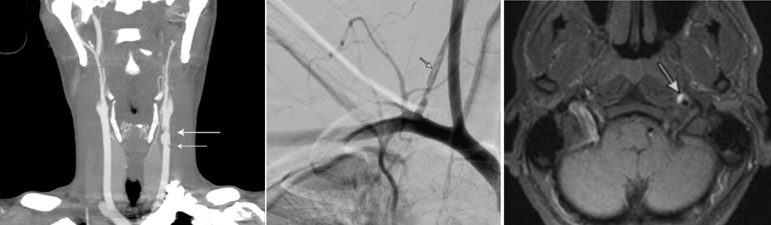
Multidetector CT angiography is the first-line imaging modality for suspected BCVI. It is rapid, widely available and can usually be incorporated into the initial trauma CT pathway. CTA should include the carotid and vertebral arteries from the aortic arch through the skull base and intracranial circulation.
Diagnostic performance depends on scanner technology, acquisition technique and image interpretation. A negative or equivocal CTA should therefore be reconsidered when the mechanism, injury pattern or clinical findings maintain a high suspicion for BCVI. This may require specialist re-review, repeat imaging or digital subtraction angiography, depending on the clinical context
Other imaging modalities
Digital subtraction angiography (DSA) remains the diagnostic reference standard but is invasive, time-consuming and not suited to routine trauma screening. It is generally reserved for equivocal or discordant CTA findings and for cases in which endovascular assessment or treatment is being considered.
Magnetic resonance angiography (MRA) avoids ionising radiation and may provide complementary vessel-wall and cerebral imaging. Longer acquisition times, limited availability and the practical difficulties of imaging unstable or multiply injured patients make it unsuitable as the primary screening test in most trauma settings
Biffl grading scale
The Biffl scale classifies BCVI according to its angiographic appearance. It standardises reporting and allows comparison on follow-up imaging, but the injury grade should be interpreted alongside symptoms, vessel involved, associated injuries and bleeding risk rather than used as an isolated treatment algorithm
| Grade | Angiographic appearance |
|---|---|
| I | Luminal irregularity, intimal dissection or intramural haematoma with <25% luminal narrowing |
| II | Dissection or intramural haematoma with ≥25% luminal narrowing, or the presence of an intraluminal thrombus or raised intimal flap |
| III | Pseudoaneurysm |
| IV | Vessel occlusion |
| V | Vessel transection with free extravasation |
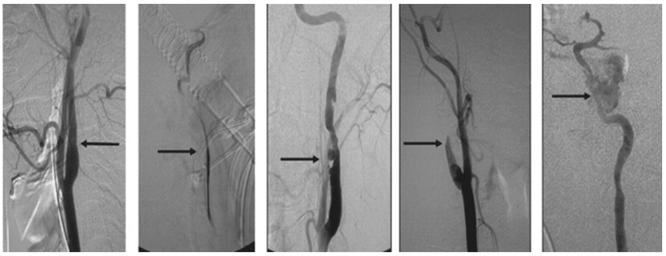
Once BCVI has been confirmed and graded, management is directed principally towards preventing thromboembolic stroke while balancing the haemorrhagic risks of associated trauma.
Management and follow-up
Start antithrombotic therapy when safe
Begin antithrombotic therapy as soon as the competing risk of traumatic haemorrhage permits. Treatment is associated with substantially lower rates of stroke and mortality than no antithrombotic treatment, although the supporting evidence is predominantly observational. The choice and timing of therapy should be individualised according to intracranial haemorrhage, solid-organ injury, operative requirements, vessel grade and local multidisciplinary practice.
Antiplatelet or anticoagulant therapy?
No randomised trial has established the optimal antithrombotic regimen for traumatic BCVI. A 2024 systematic review and meta-analysis of 39 observational studies found lower pooled rates of stroke and bleeding with antiplatelet therapy than with anticoagulation. In studies directly comparing aspirin with heparin, stroke rates were similar, while bleeding complications were less frequent with aspirin. Antiplatelet therapy is therefore a reasonable first-line strategy for many patients, but treatment should remain individualised because selection bias and clinical heterogeneity limit the certainty of the evidence
Follow-up imaging
Repeat CTA at approximately 7 days can confirm or reject the initial diagnosis, identify progression and show whether an apparent lesion represented vasospasm or imaging artefact. Antithrombotic therapy is generally continued for at least 3 months, with further imaging at around 3 months helping to guide cessation, continuation or modification of treatment.
Follow-up timing should be adapted to injury grade, symptoms and local specialist practice.
Endovascular and operative treatment
Routine stenting of grade II or III BCVI is not recommended because it has not reduced stroke compared with antithrombotic therapy alone and introduces procedural and in-stent thrombosis risks. Neurointerventional review may be appropriate for persistent or enlarging pseudoaneurysm, progressive severe stenosis with thrombotic or ischaemic complications, selected arteriovenous fistulae and injuries requiring endovascular haemorrhage control. Grade V injury requires immediate multidisciplinary management according to the site of transection, accessibility and haemodynamic state.
New or progressive neurological findings should prompt immediate reassessment, repeat vascular and cerebral imaging, and activation of the appropriate stroke and neurointerventional pathway.
Blunt Cerebrovascular Injuries in Children
Paediatric BCVI is uncommon but probably under-recognised because screening practices and imaging thresholds vary between centres. Important associated findings include skull-base fracture, cervical spine injury, intracranial haemorrhage, mandibular fracture, severe traumatic brain injury and high overall injury severity.
No paediatric screening tool has been consistently validated across populations. Adult criteria, including the Expanded Denver criteria, are commonly applied, but paediatric studies have reported variable and sometimes poor sensitivity. Decisions about CTA should therefore balance mechanism and associated injuries against radiation exposure and should involve local paediatric trauma and radiology expertise.
Treatment is generally adapted from adult practice, but the paediatric evidence base remains limited. Antiplatelet and anticoagulant therapies have both been used, with agent, timing and duration individualised according to injury grade, cerebral ischaemia, intracranial haemorrhage, associated injuries and specialist advice. The optimal regimen and follow-up schedule remain uncertain.
Peer reviewed by: Dr M Moores, stroke neurologist; Dr S Watson, stroke fellow and emergency physician; Dr S Nugent, trauma and emergency physician; and the trauma team at Kelowna General Hospital.
References
- Duke BJ, Partington MD. Blunt carotid injury in children. Pediatr Neurosurg. 1996 Oct;25(4):188-93.
- Fabian TC, Patton JH Jr, Croce MA, Minard G, Kudsk KA, Pritchard FE. Blunt carotid injury. Importance of early diagnosis and anticoagulant therapy. Ann Surg. 1996 May;223(5):513-22; discussion 522-5.
- Lew SM, Frumiento C, Wald SL. Pediatric blunt carotid injury: a review of the National Pediatric Trauma Registry. Pediatr Neurosurg. 1999 May;30(5):239-44.
- Biffl WL, Moore EE, Offner PJ, Brega KE, Franciose RJ, Burch JM. Blunt carotid arterial injuries: implications of a new grading scale. J Trauma. 1999 Nov;47(5):845-53
- Biffl WL, Ray CE Jr, Moore EE, Franciose RJ, Aly S, Heyrosa MG, Johnson JL, Burch JM. Treatment-related outcomes from blunt cerebrovascular injuries: importance of routine follow-up arteriography. Ann Surg. 2002 May;235(5):699-706; discussion 706-7.
- Rozycki GS, Tremblay L, Feliciano DV, Tchorz K, Hattaway A, Fountain J, Pettitt BJ. A prospective study for the detection of vascular injury in adult and pediatric patients with cervicothoracic seat belt signs. J Trauma. 2002 Apr;52(4):618-23; discussion 623-4.
- Miller PR, Fabian TC, Croce MA, Cagiannos C, Williams JS, Vang M, Qaisi WG, Felker RE, Timmons SD. Prospective screening for blunt cerebrovascular injuries: analysis of diagnostic modalities and outcomes. Ann Surg. 2002 Sep;236(3):386-93; discussion 393-5
- Cothren CC, Moore EE, Ray CE Jr, Ciesla DJ, Johnson JL, Moore JB, Burch JM. Screening for blunt cerebrovascular injuries is cost-effective. Am J Surg. 2005 Dec;190(6):845-9.
- Edwards NM, Fabian TC, Claridge JA, Timmons SD, Fischer PE, Croce MA. Antithrombotic therapy and endovascular stents are effective treatment for blunt carotid injuries: results from longterm followup. J Am Coll Surg. 2007 May;204(5):1007-13; discussion 1014-5.
- Tisherman SA, Bokhari F, Collier B, Cumming J, Ebert J, Holevar M, Kurek S, Leon S, Rhee P. Clinical practice guideline: penetrating zone II neck trauma. J Trauma. 2008 May;64(5):1392-405.
- Biffl WL, Cothren CC, Moore EE, Kozar R, Cocanour C, Davis JW, McIntyre RC Jr, West MA, Moore FA. Western Trauma Association critical decisions in trauma: screening for and treatment of blunt cerebrovascular injuries. J Trauma. 2009 Dec;67(6):1150-3. [PDF]
- Cothren CC, Biffl WL, Moore EE, Kashuk JL, Johnson JL. Treatment for blunt cerebrovascular injuries: equivalence of anticoagulation and antiplatelet agents. Arch Surg. 2009 Jul;144(7):685-90
- Bromberg WJ, Collier BC, Diebel LN, Dwyer KM, Holevar MR, Jacobs DG, Kurek SJ, Schreiber MA, Shapiro ML, Vogel TR. Blunt cerebrovascular injury practice management guidelines: the Eastern Association for the Surgery of Trauma. J Trauma. 2010 Feb;68(2):471-7. [Online resource]
- Ciapetti M et al. Diagnosis of Carotid arterial injury in major trauma using modification of Memphis criteria. Scand J Trauma Resusc Emerg Med 2010;18:61
- Geddes AE, Burlew CC, Wagenaar AE, Biffl WL, Johnson JL, Pieracci FM, Campion EM, Moore EE. Expanded screening criteria for blunt cerebrovascular injury: a bigger impact than anticipated. Am J Surg. 2016 Dec;212(6):1167-1174.
- Foreman PM, Griessenauer CJ, Kicielinski KP, Schmalz PGR, Rocque BG, Fusco MR, Sullivan JC 3rd, Deveikis JP, Harrigan MR. Reliability assessment of the Biffl Scale for blunt traumatic cerebrovascular injury as detected on computer tomography angiography. J Neurosurg. 2017 Jul;127(1):32-35
- Brommeland T, Helseth E, Aarhus M, Moen KG, Dyrskog S, Bergholt B, Olivecrona Z, Jeppesen E. Best practice guidelines for blunt cerebrovascular injury (BCVI). Scand J Trauma Resusc Emerg Med. 2018 Oct 29;26(1):90.
- Lauerman MH, Irizarry K, Sliker C, Bruns BR, Tesoriero R, Scalea TM, Stein DM. Influence of luminal stenosis in aneurysmal and non-aneurysmal blunt cerebrovascular injury. Injury. 2019 Jan;50(1):131-136.
- Chaibi A, Russell MB. A risk-benefit assessment strategy to exclude cervical artery dissection in spinal manual-therapy: a comprehensive review. Ann Med. 2019 Mar;51(2):118-127.
- Kim DY, Biffl W, Bokhari F, Brakenridge S, Chao E, Claridge JA, Fraser D, Jawa R, Kasotakis G, Kerwin A, Khan U, Kurek S, Plurad D, Robinson BRH, Stassen N, Tesoriero R, Yorkgitis B, Como JJ. Evaluation and management of blunt cerebrovascular injury: A practice management guideline from the Eastern Association for the Surgery of Trauma. J Trauma Acute Care Surg. 2020 Jun;88(6):875-887.
- Black JA, Abraham PJ, Abraham MN, Cox DB, Griffin RL, Holcomb JB, Hu PJ, Kerby JD, Liptrap EJ, Thaci B, Harrigan MR, Jansen JO. Universal screening for blunt cerebrovascular injury. J Trauma Acute Care Surg. 2021 Feb 1;90(2):224-231
- Yaghi S et al. Antithrombotic Treatment for Stroke Prevention in Cervical Artery Dissection: The STOP-CAD Study. Stroke. 2024 Apr;55(4):908-918
- Momic J, Yassin N, Kim MY, Walser E, Smith S, Ball I, Moffat B, Parry N, Vogt K. Antiplatelets versus anticoagulants in the treatment of blunt cerebrovascular injury (BCVI) – A systematic review and meta-analysis. Injury. 2024 Apr;55(4):111485.
Trauma Library
Injury database

Dr Neil Long BMBS FACEM FRCEM FRCPC. Emergency Physician at Kelowna hospital, British Columbia. Loves the misery of alpine climbing and working in austere environments (namely tertiary trauma centres). Supporter of FOAMed, lifelong education and trying to find that elusive peak performance.
Your posts always 💯 inspire me to think differently about these topics