


Böhler angle
Böhler angle: an acute angle measured on a lateral radiograph of the calcaneus, formed by the intersection of a line joining the posterior calcaneal tuberosity to the highest point of the posterior facet and a second line joining the posterior facet to the highest point of the anterior calcaneal process.
It provides a two-dimensional estimate of calcaneal height and posterior-facet depression. Reduction, flattening or reversal of the angle may accompany a calcaneal compression fracture, particularly a displaced intra-articular fracture.
Synonyms: Böhler’s angle; Boehler angle; Bohler angle; tuber-joint angle; tuber angle; calcaneal angle.
Definition and measurement
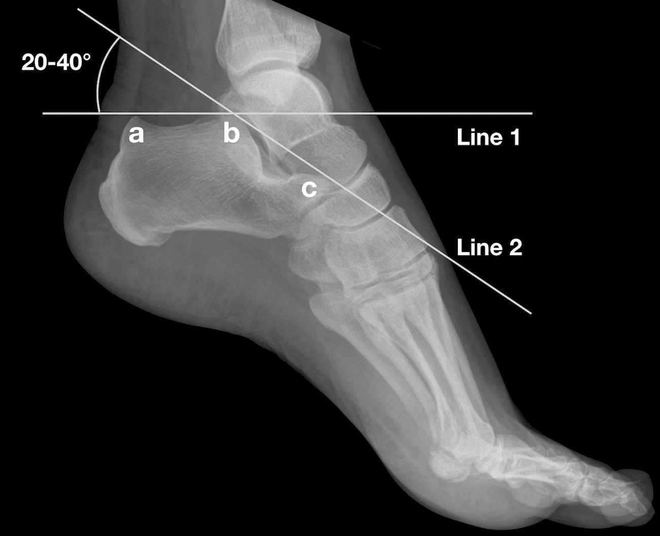
The angle is measured on a true lateral foot or calcaneal radiograph using three landmarks:
- a: highest point of the posterior calcaneal tuberosity
- b: highest point of the posterior articular facet
- c: highest point of the anterior calcaneal process
A line is drawn from a to b, and a second from b to c. The acute superior angle at b is the Böhler angle. Modern descriptions consistently use these three landmarks, although terminology for the anterior landmark varies between the anterior process and the superior calcaneal surface adjacent to the calcaneocuboid joint.
What the angle represents
The Böhler angle is principally a marker of:
- calcaneal height;
- position and depression of the posterior facet;
- flattening of the superior calcaneal contour; and
- overall compression or displacement of the calcaneal body.
During axial compression, the talus may drive the posterior facet inferiorly into the calcaneal body. This lowers the apex of the angle and produces a reduced, flat or occasionally negative measurement. The angle does not directly demonstrate the number, orientation or congruity of posterior-facet fragments; computed tomography is required to define intra-articular fracture morphology.
Böhler originally described the Gelenk-Tuberwinkel as 27–33° in his 1929 German paper. In his 1931 English-language account, he revised the normal range to 30–35°, although one of the illustrated normal calcanei measured 40°. A modern range of 20–40° or 25–40° is commonly quoted, but population studies demonstrate wider physiological variation.
Chen, Bohrer and Kelley measured a mean of 30° with a range of 14–50° in 120 normal radiographs. Using 28° as the lower limit would have classified 31% of their normal subjects as abnormal, whereas a threshold of 20° reduced the false-positive rate to 2.5%.
Willmott, Stanton and Southgate found a mean of 36.4° in 128 uninjured British adults, with a total range of 24.7–48.9° and a calculated 95% reference range of 28.2–44.5°. A Croatian population study reported a mean of 34° and range of 21–46°, while a Chinese study reported a mean of 31.6° and range of approximately 20–47°. These studies found no clinically important association with sex or laterality, but demonstrate that a single universal “normal” value is inappropriate.
20–40° is the commonly quoted reference range; an angle of 20° or less strongly supports fracture-related calcaneal collapse, but a normal angle does not exclude fracture.
Clinical use
Fracture detection
A reduced Böhler angle may draw attention to a calcaneal fracture on the initial lateral radiograph. In a 424-patient study, Isaacs and colleagues found that an angle of 20° or less provided the highest diagnostic accuracy, with measurements below 21° strongly associated with fracture in that cohort.
Its additional value should not be overstated. Knight and colleagues found that emergency physicians diagnosed calcaneal fractures from the radiographs with 97.9% accuracy before using either Böhler or Gissane measurements. Böhler angle had better diagnostic and interobserver performance than the Gissane angle, but one fracture visible only on CT was missed by all radiographic observers.
Assessment of displacement
The angle provides a rapid estimate of loss of calcaneal height and posterior-facet depression. A very low or negative angle usually reflects a more severe compression injury, although it cannot replace CT classification or direct assessment of the articular surface.
Assessment of reduction
Restoration of the angle is commonly recorded following reduction and fixation as one component of restoring calcaneal shape. It should be considered alongside:
- posterior-facet reduction on CT;
- calcaneal height, length and width;
- hindfoot varus or valgus;
- sustentacular fragment position; and
- soft-tissue condition.
Restoring the angle does not itself prove anatomical reduction of the subtalar joint.
Follow-up
Serial measurement may identify secondary loss of calcaneal height. However, observer disagreement of several degrees can create apparent change where none has occurred, so small postoperative differences should be interpreted cautiously.
Prognosis
An extremely reduced or negative presenting angle may function as a marker of initial injury severity. Patients presenting with an angle below 0° have shown poorer long-term outcomes and a greater likelihood of later subtalar fusion in some series. Other studies, however, found little or no independent correlation between postoperative angle restoration and patient-reported functional outcome.
The angle is best regarded as a descriptive and prognostic adjunct, rather than an independent treatment target.
Limitations
The principal limitations are:
- broad variation in normal anatomy;
- disagreement over the precise radiographic landmarks;
- measurement differences between observers;
- inability to define three-dimensional fracture morphology;
- reduced usefulness in bilateral injuries where contralateral comparison is unavailable;
- normal measurements in some nondisplaced, extra-articular or CT-only fractures; and
- inconsistent correlation with functional outcome.
Although radiographic positioning has often been blamed for measurement variability, experimental obliquity of up to 30° produced relatively modest changes in Böhler angle. Landmark selection and fracture comminution may therefore be more important sources of disagreement than small deviations from a true lateral projection.
In children, the angle changes during skeletal development. Clint and colleagues found excellent measurement reliability but age-related variation, particularly during the first decade; adult thresholds should not be applied uncritically to young children
Böhler angle versus Gissane angle
| Böhler angle | Gissane angle | |
|---|---|---|
| Landmarks | Posterior tuberosity, posterior facet and anterior process | Downward posterior-facet slope and upward anterior-process slope |
| Principal relationship | Calcaneal height and posterior-facet depression | Contour around the calcaneal sulcus and subtalar region |
| Fracture change | Usually reduced, flattened or reversed | May be increased, reduced, reversed or obscured |
| Reproducibility | Generally better | Frequently poor |
| Primary value | Estimate of calcaneal compression and restoration of height | Descriptive marker of disruption around the posterior facet |
The measurements describe different consequences of calcaneal injury and are complementary rather than interchangeable. Neither measurement should determine fracture management in isolation.
History of the Böhler angle
1929 – Lorenz Böhler (1885-1973) presented Behandlung der Fersenbeinbrüche at the 53rd meeting of the German Society of Surgery on April 6, 1929. The paper was published in Archiv für klinische Chirurgie later that year.
Böhler described an angle of 27–33° between a line joining the anterior calcaneus to the posterior articular surface and a second line passing along the superior aspect of the calcaneal tuberosity. He named it the Gelenk-Tuberwinkel (joint–tuberosity angle), and observed that after calcaneal fracture it became smaller, disappeared or became negative.
Serial radiographs demonstrated reduction of the angle after fracture and its restoration following treatment. The paper therefore contains the original definition, name, normal range and clinical application of the measurement now known as the Böhler angle.
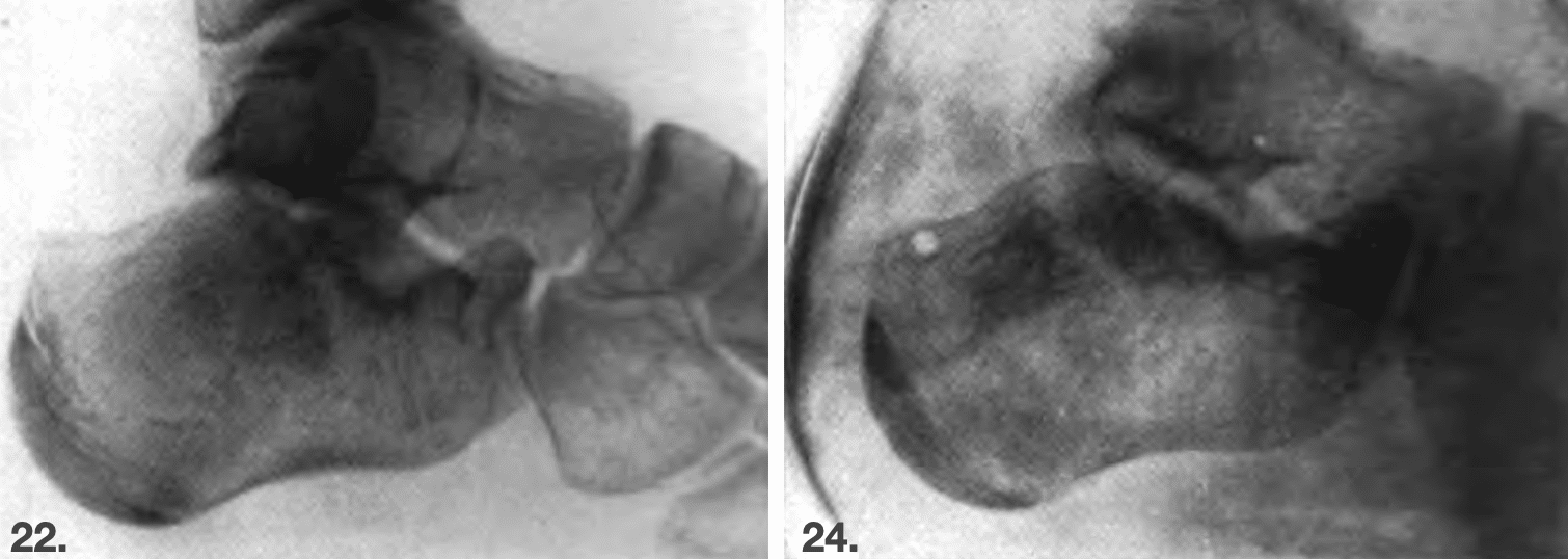
Left: lateral radiograph showing diminution of the angle with depression of the lateral posterior articular surface.
Right: follow-up radiograph three weeks after reduction showing restoration of the angle and posterior-facet position. Adapted from Böhler, 1929.
1930–1931 – Böhler delivered an invited lecture on calcaneal fractures at the American Orthopaedic Association meeting in Chatham, Massachusetts, in June 1930. The expanded English-language paper, Diagnosis, pathology and treatment of fractures of the os calcis, was published in 1931.
He translated the German Gelenk-Tuberwinkel as the tuber-joint angle and revised the stated normal range to 30–35°. The article’s illustrations of normal and fractured calcanei became the principal English-language source through which the angle entered international orthopaedic literature.
Normally there exists, between the upper contour of the tuberosity of the os calcis and the line uniting the highest point of the anterior process with the highest point of the posterior articular surface, an angle of thirty to thirty-five degrees. This angle I have named the “tuber-joint angle“. In fractures of the os calcis this angle becomes smaller, straight, or even reversed
Böhler 1931
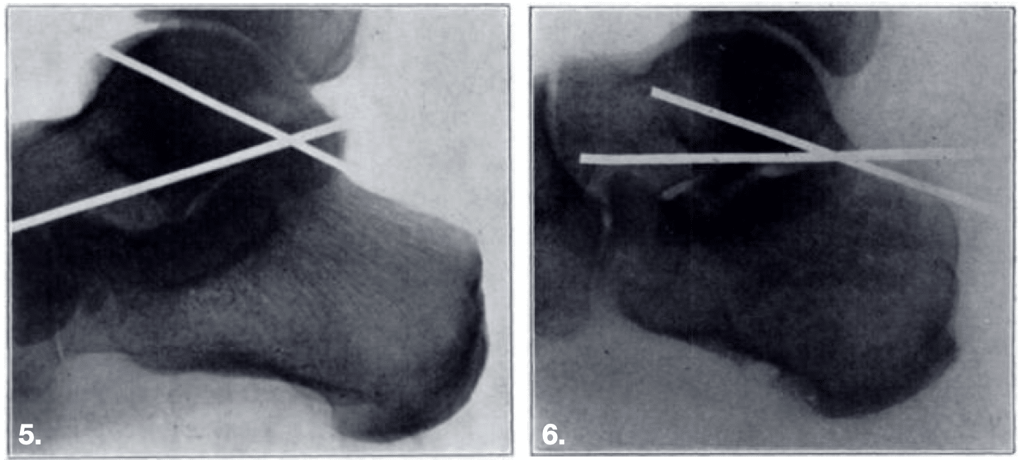
His Figure 5 illustrated a normal angle of 40° with an inclined longitudinal axis of the talus—then termed the astragalus. Figure 6 showed an angle of 20° in a mild calcaneal fracture, accompanied by flattening of the superior calcaneal contour and a more horizontal talar axis.
1975 – Hauser and Kroeker published Boehler’s angle: a review and study, one of the first papers devoted specifically to the measurement.
1991 – Chen et al measured 120 normal radiographs and found a mean of 30°, but a broad range of 14–50°. Their work demonstrated that Böhler’s original 30–35° range was too narrow for use as a strict diagnostic threshold and supported approximately 20° as a more specific lower limit.
2002 – Buckley et al, reported an association between Böhler angle and outcome in displaced intra-articular fractures. Subsequent analysis of patients requiring late subtalar fusion suggested that a presenting angle below 0° identified a particularly severe injury group at increased risk of later fusion. These studies established the angle as a marker of initial injury severity, although not necessarily an independent determinant of outcome.
2006 – Knight and colleagues compared Böhler and Gissane measurements in emergency-department radiographs. Böhler angle showed good interobserver reliability and moderate diagnostic performance, but added little to direct radiographic recognition by experienced clinicians
2007 – Schepers et al assessed radiographic measurements after displaced intra-articular fractures and found that the measured angles did not correlate with disease-specific functional outcome scores. The study reinforced the distinction between restoration of radiographic geometry and recovery of function.
2010 – Clint and colleagues found excellent interobserver and intraobserver reliability for Böhler-angle measurement in children, but demonstrated variation with skeletal development.
2012 – Willmott, Stanton and Southgate reported a mean of 36.4° and a 95% reference range of 28.2–44.5°. Despite good overall interobserver correlation, observers differed by more than 5° in approximately 41% of cases. They recommended comparison with the opposite side when the diagnosis remained uncertain.
2013 – Isaacs and colleagues compared 212 CT-confirmed fractures with 212 controls. An angle of 20° or less had the highest diagnostic accuracy in their cohort. The results support this value as a useful screening threshold, but not as an absolute rule applicable to every population or fracture pattern.
2015 – Otero and colleagues found high calculated intraclass correlations for Böhler angle but substantial absolute disagreement among surgeons when measuring displaced intra-articular fractures. Even with a wide allowable difference, consensus was absent in a significant proportion of cases.
2020 – De Boer and colleagues found that substantial experimental changes in radiographic projection altered Böhler angle by approximately 5° or less. Modest malpositioning alone therefore did not account for the wider observer disagreement reported in clinical fractures.
2021 – Nooijen et al found no significant association between six lateral radiographic measurements, including Böhler angle, and patient-reported outcomes after surgically treated intra-articular fractures. The multicentre TRON study subsequently found that loss of a restored angle was associated with greater pain, but not with a significant difference in the total AOFAS score.
2026 – Novaes and colleagues compared lateral radiographs with multiplanar CT measurements in 160 dry calcanei. Böhler angle was more reproducible than Gissane angle, with mean values of 31.8° on radiographs and 29.7° on CT. CT improved reproducibility, although the dry-bone design limits direct translation to acute clinical fractures.
Eponymic interpretation
The eponymic attribution is strong. Böhler personally defined, named and applied the measurement in his 1929 German paper, referring to it as the Gelenk-Tuberwinkel. He subsequently translated the term as tuber-joint angle and disseminated the concept internationally through his 1931 English-language publication.
Later authors replaced Böhler’s descriptive terminology with Böhler angle, while retaining essentially the same radiographic landmarks. The 1929 paper is the original description; the 1931 article is the principal English-language account.
Associated Persons
- Lorenz Böhler (1885-1973) [Böhler angle]
- William Gissane (1898-1981) [Critical angle of Gissane]
Alternative names
- Boehler’s angle, Bohler’s angle
- Tuber-joint angle, tuber angle
- Calcaneal angle
References
Historical articles
- Böhler L. Behandlung der Fersenbeinbrüche. Arch Klin Chir. 1929;157:723–732.
- Böhler L. Diagnosis, pathology and treatment of fractures of the os calcis. J Bone Joint Surg 1931; 13: 75-89
Review articles
- Hauser ML, Kroeker RO. Boehler’s angle: a review and study. J Am Podiatry Assoc. 1975 Jun;65(6):517-21.
- Chen MY, Bohrer SP, Kelley TF. Boehler’s angle: a reappraisal. Ann Emerg Med. 1991; 20(2): 122-4.
- Sanders R. Intra-articular fractures of the calcaneus: present state of the art. J Orthop Trauma. 1992; (2): 252-265.
- Buckley R et al. Operative compared with nonoperative treatment of displaced intra-articular calcaneal fractures: a prospective, randomized controlled multicenter trial. J Bone Joint Surg Am. 2002; 84: 1733-1744.
- Csizy M, Buckley R, Tough S, Leighton R, Smith J, McCormack R, Pate G, Petrie D, Galpin R. Displaced intra-articular calcaneal fractures: variables predicting late subtalar fusion. J Orthop Trauma. 2003 Feb;17(2):106-12.
- Knight JR, Gross EA, Bradley GH, Bay C, LoVecchio F. Boehler’s angle and the critical angle of Gissane are of limited use in diagnosing calcaneus fractures in the ED. The American Journal of Emergency Medicine, 2006; 24(4): 423–427
- Schepers T, Ginai AZ, Mulder PG, Patka P. Radiographic evaluation of calcaneal fractures: to measure or not to measure. Skeletal Radiol. 2007 Sep;36(9):847-52.
- Clint SA, Morris TP, Shaw OM, Oddy MJ, Rudge B, Barry M. The reliability and variation of measurements of the os calcis angles in children. J Bone Joint Surg Br. 2010 Apr;92(4):571-5
- Boyle MJ et al. The paediatric Bohler’s angle and crucial angle of Gissane: a case series. Journal of Orthopaedic Surgery and Research 2011, 6:2
- Willmott H, Stanton J, Southgate C. Böhler’s angle – What is normal in the uninjured British population? Foot Ankle Surg. 2012 Sep;18(3):187-9.
- Isaacs JD, Baba M, Huang P, Symes M, Guzman M, Nandapalan H, Moopanar T, Marchalleck S, Szomor Z. The diagnostic accuracy of Böhler’s angle in fractures of the calcaneus. J Emerg Med. 2013 Dec;45(6):879-84
- Arslan G, Yirgin IK, Tasguzen A. A measuremental approach to calcaneal fractures. Eur J Trauma Emerg Surg. 2014; 40(5): 593-9
- Nawfar SA, Chan KL, Idham HM, Izani IM, Nahulan T. Outcome Determining Factors for displaced Intra-articular Calcaneal Fractures treated operatively. Malays Orthop J. 2015 Nov;9(3):8-16
- Otero JE, Westerlind BO, Tantavisut S, Karam MD, Phisitkul P, Akoh CC, Gao Y, Marsh JL. There is poor reliability of Böhler’s angle and the crucial angle of Gissane in assessing displaced intra-articular calcaneal fractures. Foot Ankle Surg. 2015 Dec;21(4):277-81.
- De Boer AS, Van Lieshout EMM, Vellekoop L, Den Hartog D, Kleinrensink GJ, Verhofstad MHJ. The Influence of Radiograph Obliquity on Böhler’s and Gissane’s Angles in Calcanei. J Foot Ankle Surg. 2020 Jan-Feb;59(1):44-47.
- Nooijen LE, Spierings KE, Sanders FRK, Dingemans SA, Halm JA, Schepers T. Determining the Correlation Between Lateral Radiograph Morphology and the Outcome Following Surgically Treated Intra-Articular Calcaneal Fractures. Foot Ankle Spec. 2021 Apr;14(2):105-113.
- Novaes T, Casagrande E, Santos AS, Zharikov Y, Fernandes R, Pires L, Babinski M, Gameiro V. Comparison and agreement analysis of Böhler and Gissane angles on radiographs and CT in dry human calcanei. J Orthop Surg Res. 2026 Jan 30;21(1):160.
- Cadogan M. Eponymythology of foot injuries. LITFL
eponymictionary
the names behind the name

MBChB - Liverpool. RMO in Emergency Medicine at Sir Charles Gairdner Hospital, currently soaking up the sun down under. Aiming to pursue a career in anaesthetics.

BA MA (Oxon) MBChB (Edin) FACEM FFSEM. Emergency physician, Sir Charles Gairdner Hospital. Passion for rugby, medical history, medical education, and asynchronous learning #FOAMed evangelist. Editor-in-Chief and Webmaster of Life in the Fast lane, LITFL | On Call 4e| Eponyms | Books | Horology |