


Chest X-ray Essentials
Imaging
The team at Medmastery are providing LITFL readers with a series of FOAMed courses from across their website.
Description
Being able to interpret the basic findings on a chest x-ray (CXR) is one of the most important clinical skills. Whether it’s pulmonary congestion, pneumonia, pleural effusion, or cardiomyopathy, many common and life-threatening problems can be readily diagnosed with the help of a chest x-ray. In this course, you’ll learn the most essential chest x-ray interpretation skills. Come in and become an expert.
Course Details

78 Lessons

12 Quizzes

7 CME
Study the full course
Visit the Medmastery website to unlock the complete course and continue your learning journey.
Course Highlights
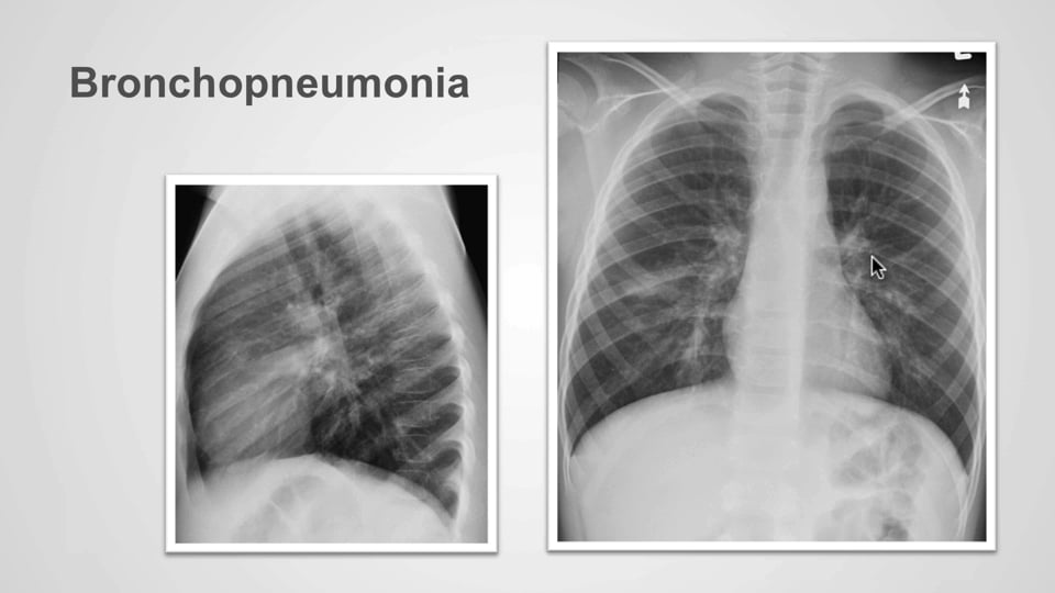
This video helps identify the various radiological presentations of pneumonia such as lobar, interstitial , bronchopneumonia and special considerations in immunocompromised patients.
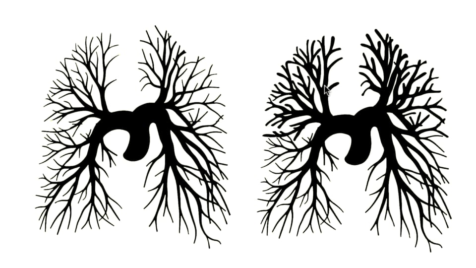
This video helps identify the features of cardiac failure on the chest X-ray. Evaluation of the three stages of failure including vascular redistribution; interstitial oedema and alveolar oedema
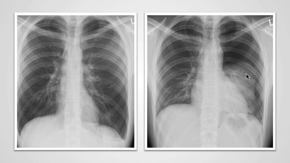
Pleural space – when the pleura is too black
This video helps you search for and identify a pneumothorax on CXR.
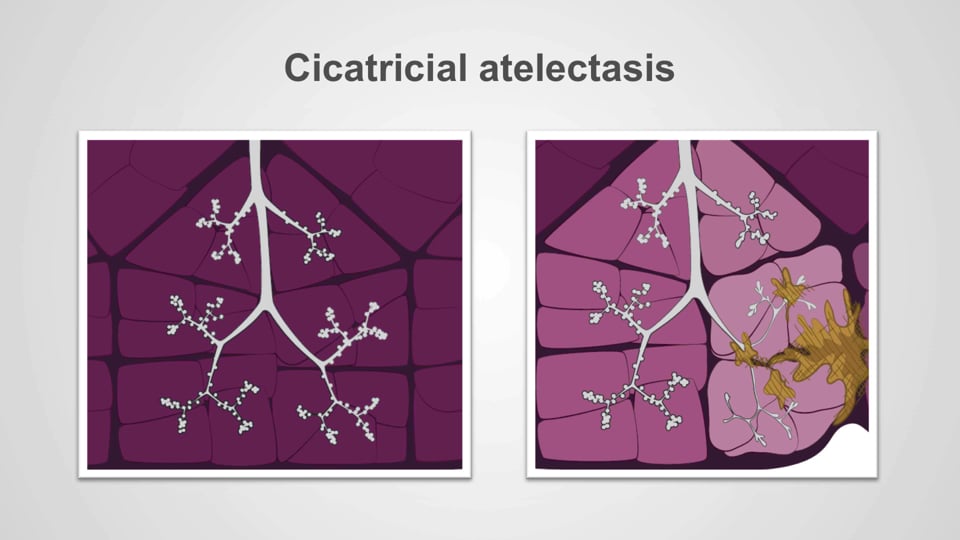
This video helps you classify and identify different types of atelectasis on Chest X-ray. Atelectasis defined as reduced inflation of all, or part, of the lung.
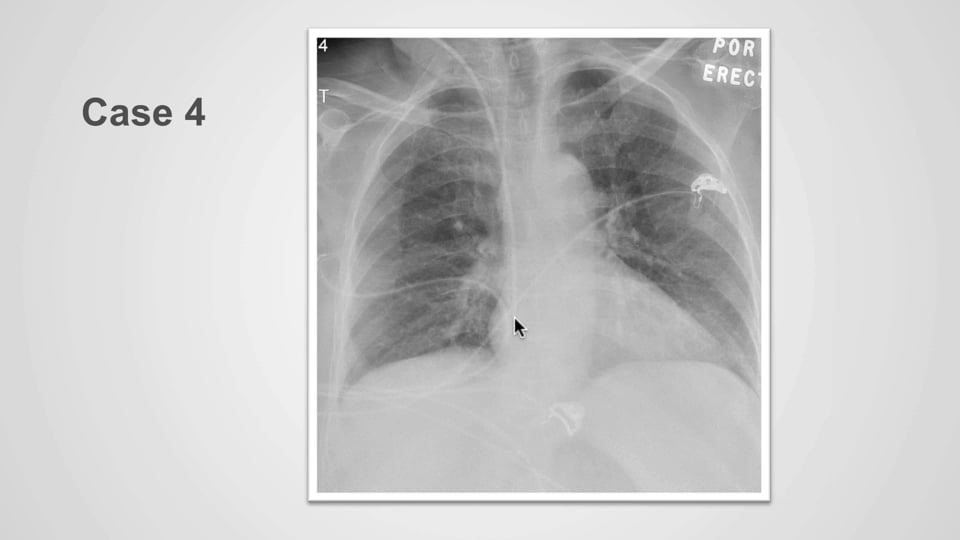
Abnormal central venous catheter positioning
This video helps identify incorrectly positioned central venous lines on chest x-ray and the potential complications of pneumothorax, pneumomediastinum, hydrothorax, hemothorax and cardiac tamponade.
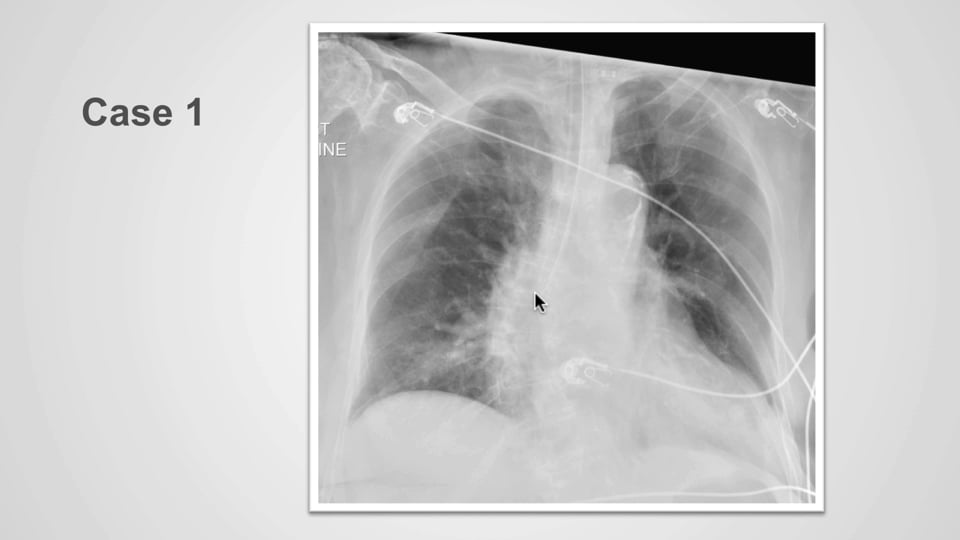
This video helps you identify aberrantly positioned endotracheal tubes (ETT) on chest x-ray by locating the tip of the ETT and the position of the carina.
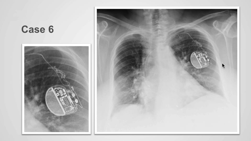
Abnormal cardiac pacer leads positioning
This video helps identify aberrantly placed cardiac pacer leads and complications such as pneumothorax, pneumomediastinum, hydrothorax, hemothorax, hydromediastinum and cardiac tamponade.