![]()
Chopart fracture dislocation
Description
Chopart Joint: articulation between the hindfoot (calcaneus and talus) and the midfoot (navicular, cuboid and cuneiforms) comprising the calcaneocuboid and talocnavicular joints. These two joints lie in a plane perpendicular to the longitudinal arch of the foot, and act as a single unit with respect to the hindfoot.
Chopart ligament: bifurcate ligament comprising the calcaneonavicular and calcaneocuboid ligaments.
Chopart line: smooth apposition of the midtarsal joint lines of the talonavicular and calcaneocuboid joints seen on both AP and lateral foot X-Rays.
Chopart fracture dislocation A dislocation of the foot through the talonavicular and the calcaneocuboid joints (Chopart joint) with associated fractures

History of the Chopart fracture dislocation
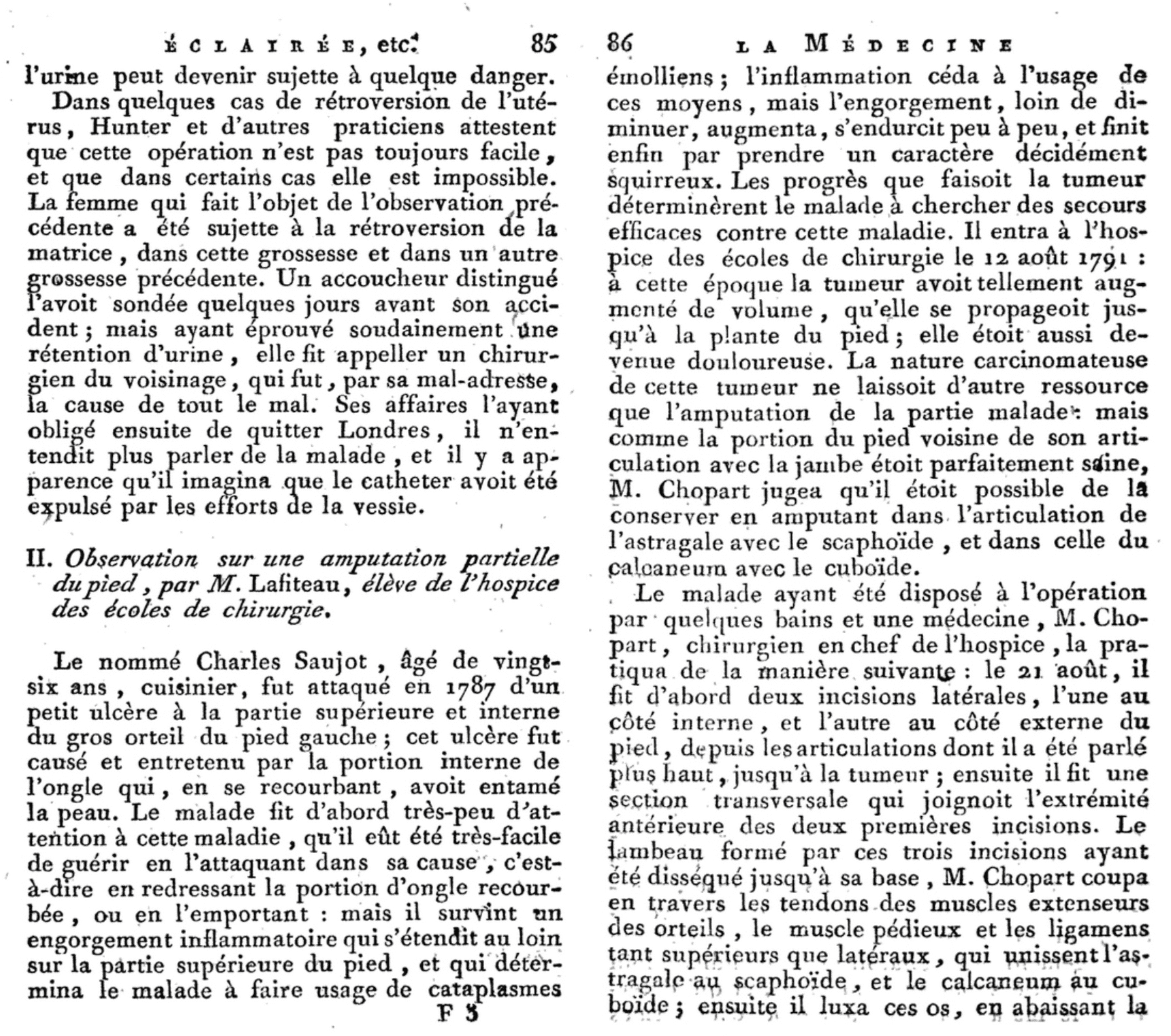
1792 – Lafiteau provided the first description of Chopart’s method of partial amputation of the foot and Chopart’s joint in Volume IV of Fourcroy ‘La médecine éclairée par les sciences physiques‘ in 1792. The fracture-dislocation was attributed at a later date.
Original
English
Charles Saujout, âgé de vingt-six ans, cuisinier, fut attaqué en 1787 d’un petit ulcère à la partie supérieure du gros orteil du pied gauche (…) il survint un engorgement inflammatoire qui s’étendit au loin sur la partie supérieure du pied (…)
La nature carcinomateuse de cette tumeur ne laissoit d’autre resource que l’amputation de la partie malade: mais comme la portion du pied voisine de son articulation avec la jambe étoit parfaitement saine, M. Chopart jugea qu’il étoit possible de la conserver en amputant dans l’articulation de l’astragale avec le scaphoïde, et dans celle du calcaneum avec le cuboïde.
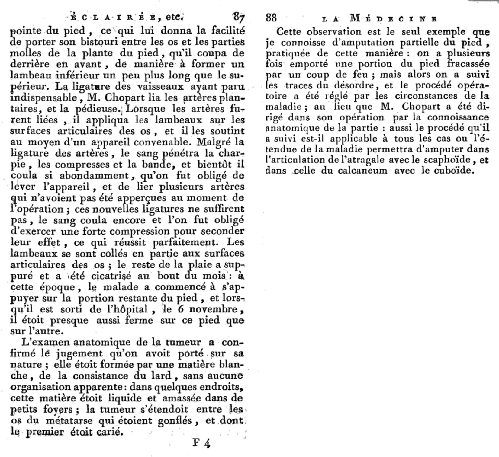
Le 21 août, il fit d’abord deux incisions latérales, l’une au côté interne et l’autre au côté externe du pied (…) ensuite il fit une section transversale qui joignait l’extrémité antérieure des deux premières incisions: le lambeau formé par ces trois incisions ayant été disséqué jusqu’à sa base, M. Chopart coupa en travers les tendons des muscles extenseurs des orteils, le muscle pédieux et les ligamens tant supérieurs que latéraux qui unissent l’astragale au scaphoïde et la calcaneum au cuboide; ensuite il luxa ces os en abaissant la pointe du pied, ce qui lui donna la facilité de porter son bistouri entre les os et le parties molles de la plante du pied qu’il coupa de derrière en devant de manière à former un lambeau inférieur un peu plus long que le supérieur.
Charles Saujout, aged 26 years, chef, suffered in 1787 a small ulcer on the top of his big toe on the left foot (…) an inflammatory engorgement ensued, which extended far up onto the superior aspect of the foot (…)
The carcinomatous nature of this tumour left no other option, but to amputate the diseased part: but since the portion of the foot next to the articulation with the leg was perfectly healthy, Mr. Chopart judged that it would be possible to conserve the former by amputating in the articulation of the talus with the navicular, and of the calcaneus with the cuboid.
On the 21st of August, he first made two lateral incisions, one on the internal, one on the external side of the foot (…) he then made a transverse incision joining the anterior extremities of the two first incisions: having dissected the flap formed by these incisions down to it’s base, Mr. Chopart transected the tendons of the extensor muscles of the toes, and the superior and lateral ligaments which bind the talus to the navicular, and the calcaneus to the cuboid; he then luxated these bones by lowering the tip of the foot, allowing him to bring his scalpel between the bones and the soft tissue of the sole of the foot, which he cut from back to front, such as to form an inferior flap slightly longer than the superior one.
Like Lisfranc (with the tarsometatarsal joint), Chopart had his procedure of midtarsal joint and the attributed fracture-dislocation injury eponymously attributed to him.
Original
English
La gloire de remettre en usage l’ablation partielle du pied dans les articulations astragalo-scaphoïdienne et calcanéo-cuboïdienne, était réservée à Chopart; d’ailleurs, les détails opératoires donnés avant lui se réduisent à rien, ou sont trop cruels pour être praticables.
The fame of reíntroducing the partial ablation of the foot in the talo-navicular and calcaneo-cuboid articulations, is attributed to Chopart; in any case, the operation reports recorded before him amount to nothing, or are too cruel to be practicable.
Associated Persons
- François Chopart (1743–1795)
Alternative names
- Chopart ligament: Bifurcate ligament
- Chopart joint: Mid-tarsal joint, transverse tarsal joint
References
Historical articles
- Lafiteau. “Observation sur une amputation partielle du pied” In: Antoine François Fourcroy. La médecine éclairée par les sciences physiques, ou Journal des découvertes relatives aux différentes parties de l’art de guéri. Paris, chez Buisoon, 1792. Volume IV p85-88. [Volume IV: 85-86; 87-88]
Reference articles
- Main BJ, Jowett RL. Injuries of the midtarsal joint. J Bone Joint Surg Br. 1975; 57(1): 89-97
- Klaue K. Chopart fractures. Injury. 2004; 35(2): SB64-70.
- Ip KY, Lui TH. Isolated dorsal midtarsal (Chopart) dislocation: a case report. J Orthop Surg (Hong Kong). 2006; 14(3): 357-9.
- Swords MP, Schramski M, Switzer K, Nemec S. Chopart fractures and dislocations. Foot Ankle Clin. 2008; 13(4): 679-93, viii.
- van Dorp KB, de Vries MR, van der Elst M, Schepers T. Chopart joint injury: a study of outcome and morbidity. J Foot Ankle Surg. 2010; 49(6): 541-5.
- Benirschke SK, Meinberg E, Anderson SA, Jones CB, Cole PA. Fractures and dislocations of the midfoot: Lisfranc and Chopart injuries. J Bone Joint Surg Am. 2012; 94(14):1325-37
- Prada A. François Chopart. 2020
- Cadogan M. Eponymous foot injuries. LITFL 2021
eponymictionary
the names behind the name

Resident medical officer in emergency medicine MB ChB (Uni. Dundee) MRCS Ed. Avid traveller, yoga teacher, polylinguist with a passion for discovering cultures.
BA MA (Oxon) MBChB (Edin) FACEM FFSEM. Emergency physician, Sir Charles Gairdner Hospital. Passion for rugby; medical history; medical education; and asynchronous learning #FOAMed evangelist. Co-founder and CTO of Life in the Fast lane | On Call: Principles and Protocol 4e| Eponyms | Books |