![]()
CT Case 089
An 88-year-old lady presents to ED with progressive decline with a background of T2DM, hypertension and hypercholesterolaemia.
She was seen four days prior at a peripheral hospital with headache and malaise. She was discharged after a reportedly normal CT brain.
Since discharge she has had increased confusion, gait disturbance and visual impairment (left homonymous hemianopia), and some subtle left sided weakness.
His CT brain is shown
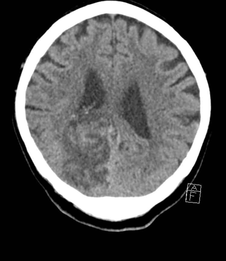
Describe and interpret the CT scans
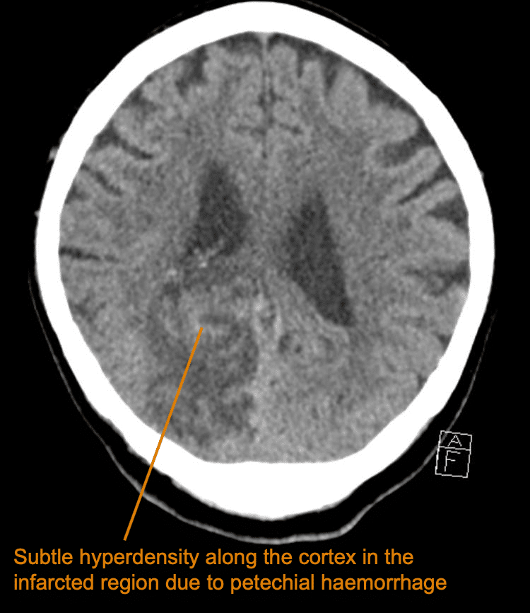
The CT demonstrates an established right PCA (posterior cerebral artery) territory infarction.
The infarct involves the cortex and the deep white matter.
There is associated oedema with mass effect which distorts the adjacent lateral ventricle.
Notice the serpiginous regions of internal hyperdensity, these represent subtle petechial haemorrhagic transformation.
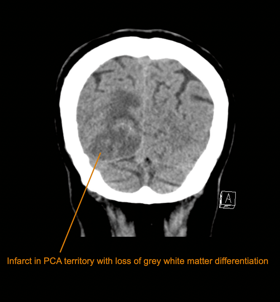
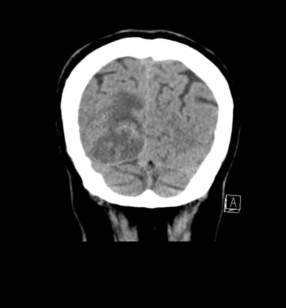
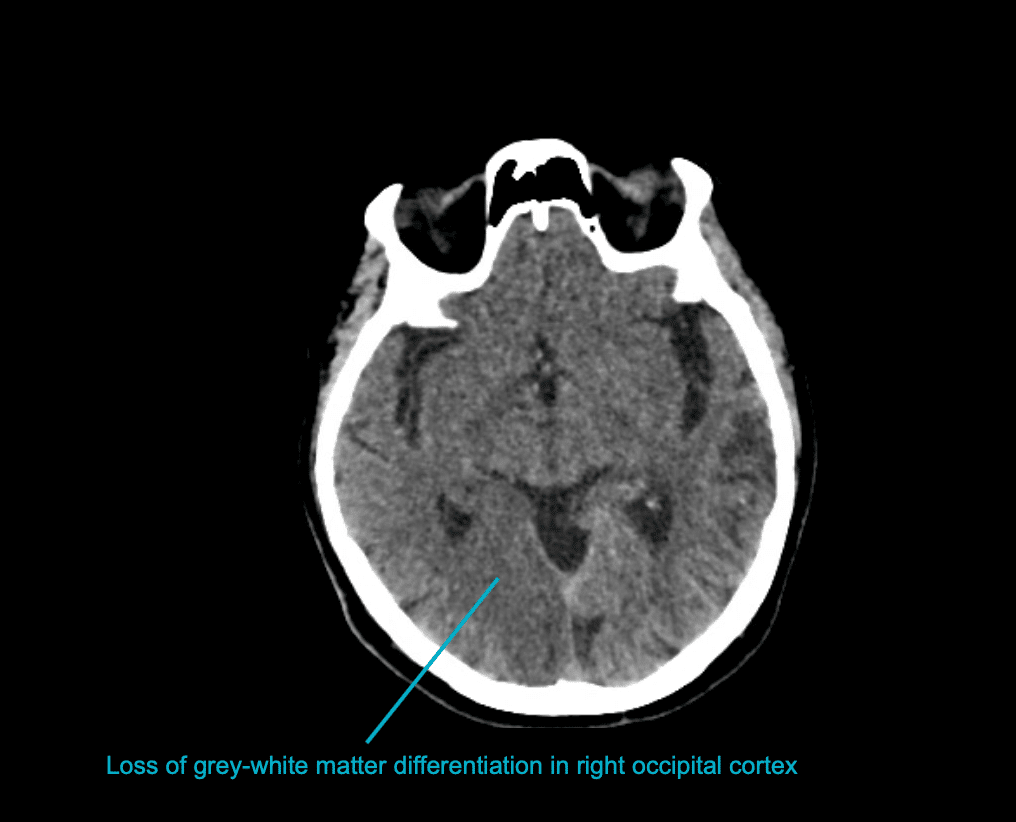
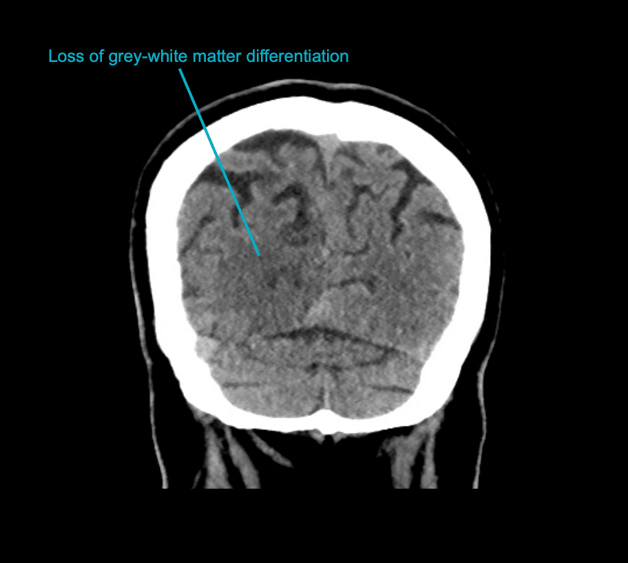
Now lets review the CT scan from 4 days earlier to see if we can identify any issues.


Describe and interpret the CT scans
While more subtle on these images, there is still evidence of infarction. We can see cytotoxic oedema with loss of great-white matter differentiation in the right occipital cortex in keeping with established ischaemic stroke.
Clinical Pearls
The extent of the PCA infarct depends upon which segment of PCA is involved.
A more proximal thrombus causes infarction of the thalamus, midbrain and cortex, while a more distal thrombus causes only cortical infarction.
This patient’s CT scan also demonstrates haemorrhagic transformation.
There are two distinct forms of haemorrhagic transformation, these are;
- Petechial haemorrhages (haemorrhagic infarction)
- Parenchymal haematoma (secondary haematoma)
Distinguishing which is present is important as each has distinct clinical and prognostic implications.
Petechial haemorrhages are due to small amounts of blood leaking out of vessels damaged by ischaemic into surrounding tissues.
Petechial haemorrhages are small and appear as subtle hyperdensities of the cortex, as seen in this case. Petechial haemorrhages do not have mass effect or impact on prognosis and treatment.
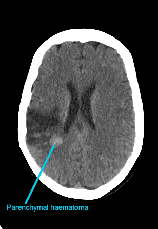
Conversely, parenchymal haematomas are far more concerning. They are larger and can cause sudden clinical deterioration due to significant mass effect. Haematomas usually appear a few days after the infarct onset. They appear as hyperdense haematomas within infarcted hypodense brain parenchyma.
Parenchymal haematoma can occur in any patient following an ischaemic stroke, however as would be expected, administration of intravenous thrombolysis significantly increases the risk.
Bonus image
This is a CT scan of a different patient it demonstrates the appearance of a parenchymal haematoma following an MCA ischaemic stroke.
TOP 100 CT SERIES

Sydney-based Emergency Physician (MBBS, FACEM) working at Liverpool Hospital. Passionate about education, trainees and travel. Special interests include radiology, orthopaedics and trauma. Creator of the Sydney Emergency XRay interpretation day (SEXI).

Provisional fellow in emergency radiology, Liverpool hospital, Sydney. Other areas of interest include paediatric and cardiac imaging.

Emergency Medicine Education Fellow at Liverpool Hospital NSW. MBBS (Hons) Monash University. Interests in indigenous health and medical education. When not in the emergency department, can most likely be found running up some mountain training for the next ultramarathon.

Dr Leon Lam FRANZCR MBBS BSci(Med). Clinical Radiologist and Senior Staff Specialist at Liverpool Hospital, Sydney