![]()
Echo basics: Aortic Valve
Mastering normal aortic valve echo anatomy
A normal aortic valve is trileaflet, with equally sized cusps that are supported by a fibrous annulus and separated by three commissures. The cusps are semilunar, or crescent-shaped, with an upward curving free edge. The slightly thickened tip is referred to as the node of Arantius
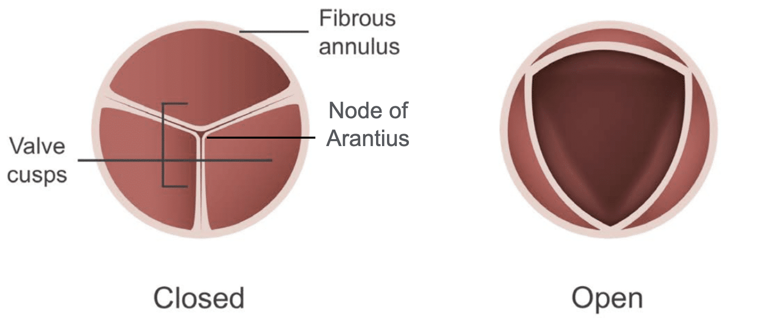
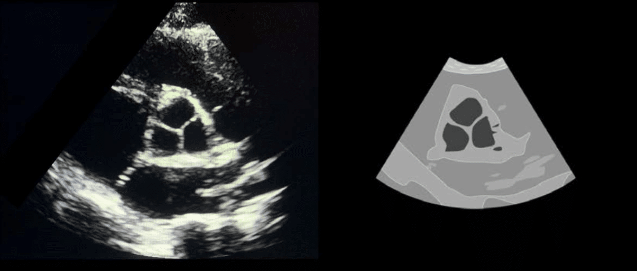
During diastole (when the valve is closed) the three nodes from each cusp meet in the centre with the closure lines forming a Y shape, when viewed from the short-axis approach.
Sometimes, thin, filament-like structures might be seen on the ventricular side of the valve cusps. These are known as Lambl’s excrescences and are generally thought to be a normal variant; however, they may arise due to gradual degeneration of the cusps. It is important to bear this feature in mind, as they might be mistaken for vegetations in an otherwise well individual.
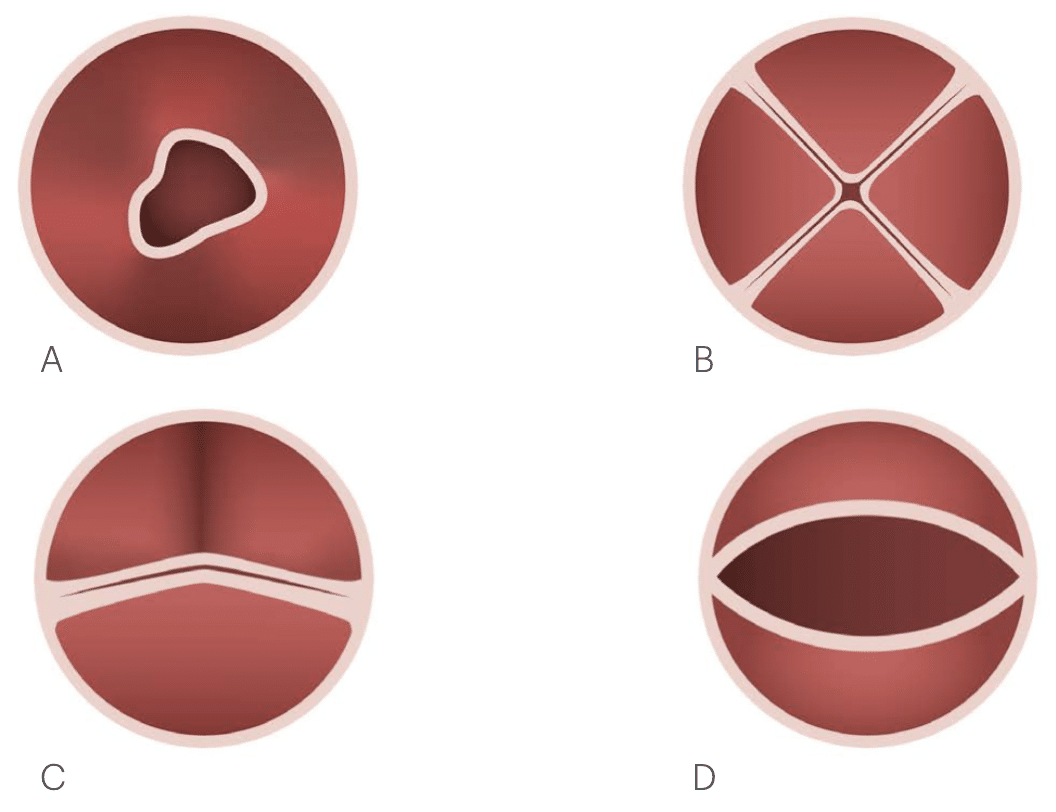
Bicuspid valves are a congenital malformation occurring in 1–2% of the population. Rarely, unicuspid and quadricuspid valves can be discovered. Abnormal cusp formation can result in valvular stenosis and regurgitation in later life.
Where the proximal aortic root meets the left ventricular outflow tract, there are three bulges that provide support for the corresponding aortic valve leaflets. These are the sinuses of Valsalva, and are named according to the origin of the coronary arteries. The right and left sinuses give rise to their respective coronary arteries, the third sinus is referred to as the noncoronary sinus, as there is no associated artery.
The sinotubular junction is the point at which the sinuses narrow and join the proximal tubular portion of the ascending aorta. When viewed in long-axis, a normal aortic valve will be seen to open rapidly as thin structures that lie parallel to the aortic walls during systole.
This is an edited excerpt from the Medmastery course Echo Masterclass – The Valves by Chris Eggett, PhD. Acknowledgement and attribution to Medmastery for providing course transcripts.
Additional echocardiography resources:
- Na, M. Echo Masterclass: Left Ventricular Strain. Medmastery
- Monteiro, C. Echo Masterclass: The Right Heart. Medmastery
- West, C. Echo Masterclass: Adult Congenital Heart Disease. Medmastery
- Naderi, H. Echo Masterclass: The Power of 3D Imaging. Medmastery
Radiology Library: Echocardiography basics
- Eggett C. Echo basics: Valve Views. LITFL
- Eggett C. Echo basics: Valves, Measurements and Reports. LITFL
- Eggett C. Echo basics: Mitral valve. LITFL
- Eggett C. Echo basics: Mitral Regurgitation. LITFL
- Eggett C. Echo basics: Mitral Stenosis. LITFL
- Eggett C. Echo basics: Aortic Valve. LITFL
- Eggett C. Echo basics: Aortic Stenosis. LITFL
- Eggett C. Echo basics: Aortic Regurgitation. LITFL
- Eggett C. Echo basics: Tricuspid Valve. LITFL
- Eggett C. Echo basics: Pulmonary Valve. LITFL
- Eggett C. Echo basics: Prosthetic Valves. LITFL
Further reading
- Otto CM. Textbook of Clinical Echocardiography. Elsevier. 7e, 2023.
- Houghton AR. Making Sense of Echocardiography: A Hands-on Guide. 3e 2023
- Otto CM, Nishimura RA, Bonow RO, Carabello BA, Erwin JP 3rd, Gentile F, Jneid H, Krieger EV, Mack M, McLeod C, O’Gara PT, Rigolin VH, Sundt TM 3rd, Thompson A, Toly C. 2020 ACC/AHA Guideline for the Management of Patients With Valvular Heart Disease: A Report of the American College of Cardiology/American Heart Association Joint Committee on Clinical Practice Guidelines. Circulation. 2021 Feb 2;143(5):e72-e227.
Echocardiography Essentials

Cardiac physiologist, echocardiographer, and Professor of Healthcare Science Education, Faculty of Medical Sciences at the University of Newcastle, UK. I direct post-grad programs at the Faculty of Medical Sciences, run an echo clinic at the Freeman Hospital, and teach transthoracic echocardiography to specialists in critical and emergency care and anaesthetic settings